

Overcoming Radiation Resistance by Iron-Platinum Metal Alloy Nanoparticles in Human Copper Transport 1-Overexpressing Cancer Cells via Mitochondrial Disturbance
- PMID: 33727814
- PMCID: PMC7955785
- DOI: 10.2147/IJN.S283147
Overcoming Radiation Resistance by Iron-Platinum Metal Alloy Nanoparticles in Human Copper Transport 1-Overexpressing Cancer Cells via Mitochondrial Disturbance
Abstract
Background: Radiation therapy remains an important treatment modality in cancer therapy, however, resistance is a major problem for treatment failure. Elevated expression of glutathione is known to associate with radiation resistance. We used glutathione overexpressing small cell lung cancer cell lines, SR3A-13 and SR3A-14, established by transfection with γ-glutamylcysteine synthetase (γ-GCS) cDNA, as a model for investigating strategies of overcoming radiation resistance. These radiation-resistant cells exhibit upregulated human copper transporter 1 (hCtr1), which also transports cisplatin. This study was initiated to investigate the effect and the underlying mechanism of iron-platinum nanoparticles (FePt NPs) on radiation sensitization in cancer cells.
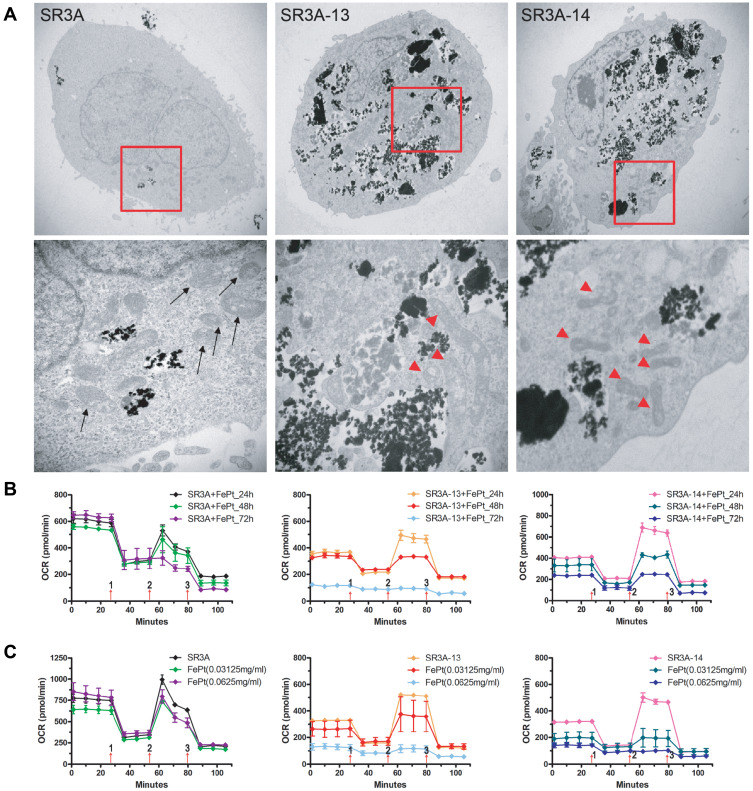
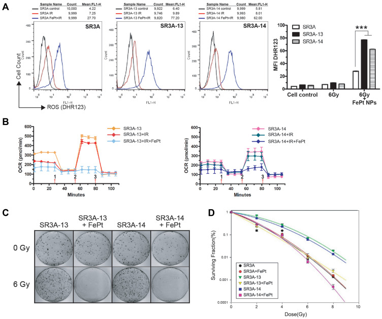
Materials and methods: Uptakes of FePt NPs in these cells were studied by plasma optical emission spectrometry and transmission electron microscopy. Effects of the combination of FePt NPs and ionizing radiation were investigated by colony formation assay and animal experiment. Intracellular reactive oxygen species (ROS) were assessed by using fluorescent probes and imaged by a fluorescence-activated-cell-sorting caliber flow cytometer. Oxygen consumption rate (OCR) in mitochondria after FePt NP and IR treatment was investigated by a Seahorse XF24 cell energy metabolism analyzer.
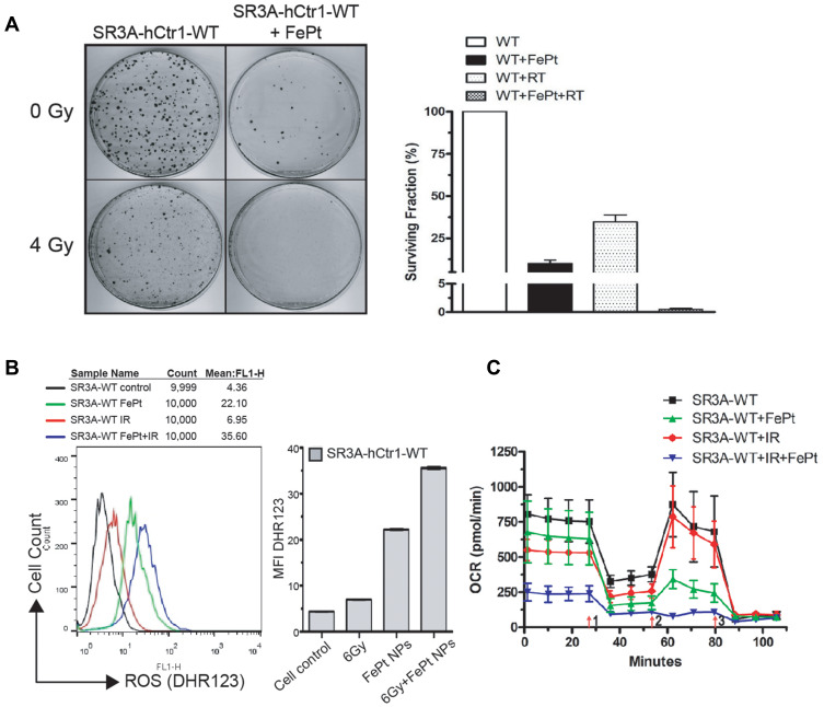
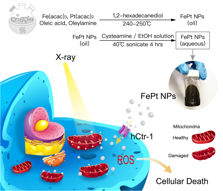
Results: These hCtr1-overexpressing cells exhibited elevated resistance to IR and the resistance could be overcome by FePt NPs via enhanced uptake of FePt NPs. Overexpression of hCtr1 was responsible for the increased uptake/transport of FePt NPs as demonstrated by using hCtr1-transfected parental SR3A (SR3A-hCtr1-WT) cells. Increased ROS and drastic mitochondrial damages with substantial reduction of oxygen consumption rate were observed in FePt NPs and IR-treated cells, indicating that structural and functional insults of mitochondria are the lethal mechanism of FePt NPs. Furthermore, FePt NPs also increased the efficacy of radiotherapy in mice bearing SR3A-hCtr1-WT-xenograft tumors.
Conclusion: These results suggest that FePt NPs can potentially be a novel strategy to improve radiotherapeutic efficacy in hCtr1-overexpressing cancer cells via enhanced uptake and mitochondria targeting.
Keywords: FePt nanoparticles; human copper transporter 1; mitochondrial targeting; radiation resistance; reactive oxygen species.
© 2021 Tsai et al.
Conflict of interest statement
The authors report no conflicts of interest in this work.
Figures


Similar articles
-
CD44-hyaluronan mediating endocytosis of iron-platinum alloy nanoparticles induces ferroptotic cell death in mesenchymal-state lung cancer cells with tyrosine kinase inhibitor resistance.Acta Biomater. 2024 Sep 15;186:396-410. doi: 10.1016/j.actbio.2024.07.020. Epub 2024 Jul 26. Acta Biomater. 2024. PMID: 39067646
-
FePt nanoparticles as a potential X-ray activated chemotherapy agent for HeLa cells.Int J Nanomedicine. 2015 Oct 22;10:6435-44. doi: 10.2147/IJN.S88458. eCollection 2015. Int J Nanomedicine. 2015. PMID: 26604740 Free PMC article.
-
Mechanistic basis for overcoming platinum resistance using copper chelating agents.Mol Cancer Ther. 2012 Nov;11(11):2483-94. doi: 10.1158/1535-7163.MCT-12-0580. Epub 2012 Aug 21. Mol Cancer Ther. 2012. PMID: 22914438 Free PMC article.
-
Overcoming platinum drug resistance with copper-lowering agents.Anticancer Res. 2013 Oct;33(10):4157-61. Anticancer Res. 2013. PMID: 24122978 Free PMC article. Review.
-
Role of the human high-affinity copper transporter in copper homeostasis regulation and cisplatin sensitivity in cancer chemotherapy.Cancer Res. 2012 Sep 15;72(18):4616-21. doi: 10.1158/0008-5472.CAN-12-0888. Epub 2012 Sep 7. Cancer Res. 2012. PMID: 22962276 Free PMC article. Review.
Cited by
-
Nanoparticles augment the therapeutic window of RT and immunotherapy for treating cancers: pivotal role of autophagy.Theranostics. 2023 Jan 1;13(1):40-58. doi: 10.7150/thno.77233. eCollection 2023. Theranostics. 2023. PMID: 36593951 Free PMC article. Review.
-
Correlation study of LINC02609 and SNHG17 as prognostic biomarkers of kidney renal clear cell carcinoma and therapeutic sensitivity based on public data and In Vitro analysis.Front Immunol. 2025 May 26;16:1592474. doi: 10.3389/fimmu.2025.1592474. eCollection 2025. Front Immunol. 2025. PMID: 40491925 Free PMC article.
-
IR808-ATIPA: A Dual-Function Agent for Enhanced Computed Tomography Imaging and Radiotherapy Sensitization in Cervical Cancer Treatment.Biomater Res. 2025 Aug 18;29:0222. doi: 10.34133/bmr.0222. eCollection 2025. Biomater Res. 2025. PMID: 40831788 Free PMC article.
-
Green Synthesis of Platinum Nanoparticles for Biomedical Applications.J Funct Biomater. 2022 Nov 21;13(4):260. doi: 10.3390/jfb13040260. J Funct Biomater. 2022. PMID: 36412901 Free PMC article. Review.
-
Application of Nanoparticles in the Diagnosis and Treatment of Colorectal Cancer.Anticancer Agents Med Chem. 2024;24(18):1305-1326. doi: 10.2174/0118715206323900240807110122. Anticancer Agents Med Chem. 2024. PMID: 39129164 Free PMC article. Review.
References
-
- Baumann M, Krause M, Overgaard J, et al. Radiation oncology in the era of precision medicine. Nat Rev Cancer. 2016;16:234–249. - PubMed
MeSH terms
Substances
LinkOut - more resources
Full Text Sources
Other Literature Sources
Medical
Research Materials
Miscellaneous
