

A case of bulla formation after treatment for COVID-19 pneumonia
- PMID: 33728014
- PMCID: PMC7945865
- DOI: 10.1016/j.radcr.2021.03.003
A case of bulla formation after treatment for COVID-19 pneumonia
Erratum in
-
Erratum regarding missing Declaration of Competing Interest statements in previously published articles.Radiol Case Rep. 2022 Sep 29;17(12):4933. doi: 10.1016/j.radcr.2022.08.054. eCollection 2022 Dec. Radiol Case Rep. 2022. PMID: 36311872 Free PMC article.
Abstract
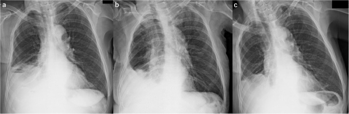
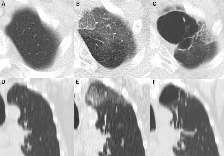
In December 2019, coronavirus disease 2019 (COVID-19), which is caused by severe acute respiratory syndrome coronavirus 2 (SARS-CoV-2), was reported in Wuhan, China. An 82-year-old woman presented to our hospital with high fever (39°C) and chest computed tomography revealed ground-glass opacities in the left lung apex. She was positive for SARS-CoV-2 based on a polymerase chain reaction test, and diagnosed with COVID-19 pneumonia. 6 months after treatment, chest CT showed a large bulla (47 mm × 29 mm) in the left lung apex, although pneumonia had partially resolved. Radiologic follow-up is needed after COVID-19 pneumonia, because patients may develop bullae after treatment.
Keywords: Bulla; COVID-19; Coronavirus; Pneumonia; SARS-CoV-2.
© 2021 The Authors. Published by Elsevier Inc. on behalf of University of Washington.
Figures
References
-
- Wiese ER. Bulla of the lung. Dis Chest. 1946;12:238–241. - PubMed
Publication types
LinkOut - more resources
Full Text Sources
Other Literature Sources
Medical
Miscellaneous
