. 2021 Feb 25:2021:2045259.
doi: 10.1155/2021/2045259. eCollection 2021.

PPAR α Targeting GDF11 Inhibits Vascular Endothelial Cell Senescence in an Atherosclerosis Model

Affiliations

PPAR α Targeting GDF11 Inhibits Vascular Endothelial Cell Senescence in an Atherosclerosis Model

Fangfang Dou et al. Oxid Med Cell Longev. .

Abstract

Atherosclerosis (AS) is a complex vascular disease that seriously harms the health of the elderly. It is closely related to endothelial cell aging, but the role of senescent cells in atherogenesis remains unclear. Studies have shown that peroxisome proliferator-activated receptor alpha (PPARα) inhibits the development of AS by regulating lipid metabolism. Our previous research showed that PPARα was involved in regulating the repair of damaged vascular endothelial cells. Using molecular biology and cell biology approaches to detect senescent cells in atherosclerosis-prone apolipoprotein E-deficient (Apoe -/-) mice, we found that PPARα delayed atherosclerotic plaque formation by inhibiting vascular endothelial cell senescence, which was achieved by regulating the expression of growth differentiation factor 11 (GDF11). GDF11 levels declined with age in several organs including the myocardium, bone, central nervous system, liver, and spleen in mice and participated in the regulation of aging. Our results showed that PPARα inhibited vascular endothelial cell senescence and apoptosis and promoted vascular endothelial cell proliferation and angiogenesis by increasing GDF11 production. Taken together, these results demonstrated that PPARα inhibited vascular endothelial cell aging by promoting the expression of the aging-related protein GDF11, thereby delaying the occurrence of AS.


Conflict of interest statement

The authors declare no conflicts of interest.

Figures
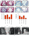
Figure 1
Figure 1
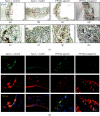
PPARα inhibits atherosclerotic plaque formation and vascular endothelial injury. (a) HE staining showed that PPARα agonists inhibited AS plaque formation. (b) Masson's trichrome staining showed that PPARα agonists promoted plaque stability in the Apoe−/− mouse model. (c) The PPARα agonist made the surface of aortic plaque covered with intact endothelial cells and inhibited endothelial cell apoptosis and necrosis. , #P < 0.05, ##P < 0.01, ∗: vs. control, #: vs. model. Scale bar, 5 μm or 10 μm or 20 μm.
Figure 2
Figure 2
PPARα inhibits aging of endothelial cells in the Apoe−/− mouse model. (a) β-gal staining showed that the number of senescent cells decreased after administration of the PPARα agonist. (b) Immunofluorescence double labeling staining showed that there were a large number of aging endothelial cells in the atherosclerotic plaque of Apoe−/− mice, and the PPARα agonist inhibited the denudation and maintained the integrity of aging endothelial cells in Apoe−/− model mice. Scale bar, 25 μm or 100 μm.
Figure 3
Figure 3
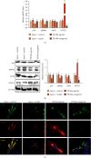
Expression of aging-related proteins in the Apoe−/− mouse aorta. (a) Real-time PCR showed that the PPARα agonist significantly inhibited the expression of p16 and p66shc and promoted the expression of GDF11 at the mRNA level, while the PPARα antagonist inhibited the expression of GDF11 at the mRNA level. (b) Western blot showed that the PPARα agonist significantly inhibited the expression of p16 and p66shc and promoted the expression of GDF11 at the protein level. In contrast, the PPARα antagonist inhibited the protein level of JunD and GDF11. (c) Immunofluorescence double labeling staining showed that the PPARα agonist promoted the expression of GDF11, while the PPARα antagonist inhibited the expression of GDF11 in vascular endothelial cells. #P < 0.05, #: vs. model. Scale bar, 100 μm.
Figure 4
Figure 4
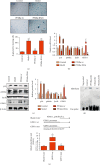
PPARα targets GDF11 expression. (a) β-gal staining showed that overexpression of PPARα significantly inhibited cell senescence. (b) Real-time PCR showed that PPARα significantly inhibited the upregulation of p16 and the downregulation of GDF11 caused by ox-LDL exposure of cultured vascular endothelial cells (200 μg/mL). (c) Western blot showed that the PPARα agonist significantly promoted GDF11 expression and inhibited p16 and p66shc expression. (d) EMSA results showed that PPARα bound to the labeled target probe, and partially to the mutant probe, indicating that PPARα could bind to the GDF11 target sequence and participate in the regulation of downstream gene transcription. (e) Double-luciferase reporter gene experiment results indicated that PPARα could bind to the GDF11-specific sequence. , #P < 0.05, ∗∗, ##P < 0.01, ∗: vs. control, #: vs. model.
Figure 5
Figure 5
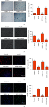
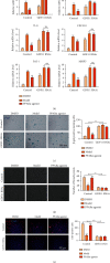
The effect of GDF11 on endothelial cell function. (a) β-gal staining showed that GDF11 inhibited vascular endothelial cell aging. (b) Angiogenesis assay showed that GDF11 improved vascular endothelial cell angiogenesis. (c) ki67 staining showed that GDF11 improved vascular endothelial cell proliferation. (d) TUNEL staining showed that GDF11 inhibited vascular endothelial cell apoptosis. , #P < 0.05, ∗: vs. control, #: vs. model. Scale bar, 100 μm or 200 μm.
Figure 6
Figure 6
PPARα influence on the function of vascular endothelial cells is achieved by regulating GDF11. (a) Western blot showed GDF11 expression while adding ox-LDL or PPARα agonist to the stable GDF11-knockdown HUVECs. (b) Real-time PCR revealed a stark increment in SASP (including TNFα, IL-1β, IL-6, CXCL10, PAI-1, and MMP3) in HUVECs (with or without GDF11 knockdown) treated with ox-LDL. (c–f) When GDF11 expression was inhibited, the ability of PPARα to promote HUVEC proliferation and angiogenesis was significantly reduced, while senescent and apoptotic cells increased significantly. ∗∗, ##P < 0.01, ∗: vs. control, #: vs. model. Scale bar, 100 μm or 200 μm.

Similar articles

Cited by

References

    1. Dong Y., Fernandes C., Liu Y., et al. Role of endoplasmic reticulum stress signalling in diabetic endothelial dysfunction and atherosclerosis. Diabetes & Vascular Disease Research. 2017;14(1):14–23. doi: 10.1177/1479164116666762. - DOI - PMC - PubMed
    1. Choi B. J., Prasad A., Gulati R., et al. Coronary endothelial dysfunction in patients with early coronary artery disease is associated with the increase in intravascular lipid core plaque. European Heart Journal. 2013;34(27):2047–2054. doi: 10.1093/eurheartj/eht132. - DOI - PMC - PubMed
    1. Erusalimsky J. D., Skene C. Mechanisms of endothelial senescence. Experimental Physiology. 2009;94(3):299–304. doi: 10.1113/expphysiol.2008.043133. - DOI - PubMed
    1. Hayashi T., Yano K., Matsui-Hirai H., Yokoo H., Hattori Y., Iguchi A. Nitric oxide and endothelial cellular senescence. Pharmacology & Therapeutics. 2008;120(3):333–339. doi: 10.1016/j.pharmthera.2008.09.002. - DOI - PubMed
    1. Lubrano V., Balzan S. LOX-1 and ROS, inseparable factors in the process of endothelial damage. Free Radical Research. 2014;48(8):841–848. doi: 10.3109/10715762.2014.929122. - DOI - PubMed

MeSH terms