Case Reports
. 2021 Feb 26:2021:6617370.
doi: 10.1155/2021/6617370. eCollection 2021.

A Case of Myoepithelial Hamartoma: Morphological Variation Supported by OCT4 Expression

Affiliations
Case Reports

A Case of Myoepithelial Hamartoma: Morphological Variation Supported by OCT4 Expression

Takehiro Tanaka et al. Case Rep Gastrointest Med. .

Abstract

In this report, we describe a patient with myoepithelial hamartoma, which is regarded as synonymous with adenomyosis and heterotopic pancreas. Endoscopy revealed a submucosal tumor in the antrum of the stomach. Subsequently, distal gastrectomy with Roux-en-Y reconstruction was performed. Histological findings of adenomyomatous lesion and heterotopic pancreatic tissue were observed in this lesion. The distribution of OCT4, which is a pluripotency marker, varied in each part.


Conflict of interest statement

The authors declare that they have no conflicts of interest.

Figures

Figure 1
Figure 1
(a) Esophagogastroduodenoscopy (EGD) image. EGF revealed a pedunculated mass resembling a submucosal tumor in the antrum of the stomach. (b) Endoscopic ultrasonography image. The lesion was located in the submucosa and muscularis propria, and the mass was poorly defined and hypoechogenic.
Figure 2
Figure 2
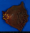
Macroscopic findings of the distal gastrectomy specimen. The lesion was located in the gastric antrum, and duodenum and was 3.7 cm long.
Figure 3
Figure 3
(a) Macroscopic findings. The resected specimen showed a vague nodule with dilated glands surrounded by edematous stroma in the submucosa and muscularis propria. (b) Histological findings. (A) Its appearance was similar to that of uterine adenomyosis. (B) The mass was composed of several types of glands, acini, and smooth muscle bundles.
Figure 4
Figure 4
Histological findings. OCT 4 is not identified in the cells of the dilated glands ((a): HE staining; (b) OCT4). A small population of small gland cells was positive for OCT4 ((c): HE staining; (d): OCT4).
Figure 5
Figure 5
Histological findings. Organoid structure composed of large ducts surrounded by radially extending acini and small ducts resembling Brunner or pyloric glands (hematoxylin and eosin staining: HE (a)). Immunohistochemical study shows that MUC5AC is negative (b), whereas MUC6 is positive (c). Acinar cells are positive for trypsin (d).

References

    1. Magnus-Alsleben E. Adenomyome des pylorus. Virchows Archiv für Pathologische Anatomie und Physiologie und für Klinische Medizin. 1903;173(1):137–155. doi: 10.1007/bf01947878. - DOI
    1. Ikegami R., Watanabe Y., Tainaka T. Myoepithelial hamartoma causing small-bowel intussusception: a case report and literature review. Pediatric Surgery International. 2005;22:387–389. - PubMed
    1. Trifan A., Târcoveanu E., Danciu M., Huţanaşu C., Cojocariu C., Stanciu C. Gastric heterotopic pancreas: an unusual case and review of the literature. Journal of Gastrointestinal and Liver Diseases: JGLD. 2012;21(2):209–212. - PubMed
    1. Zarling E. J. Gastric adenomyoma with coincidental pancreatic rest: a case report. Gastrointestinal Endoscopy. 1981;27(3):175–177. doi: 10.1016/s0016-5107(81)73186-9. - DOI - PubMed
    1. Ryan A., Lafnitzegger J. R., Lin D. H., Jakate S., Staren E. D. Myoepithelial hamartoma of the duodenal wall. Virchows Archiv. 1998;432(2):191–194. doi: 10.1007/s004280050155. - DOI - PubMed

Publication types

LinkOut - more resources