. 2021 Mar 1:2021:6694392.
doi: 10.1155/2021/6694392. eCollection 2021.

Increased Nuclear Transporter KPNA2 Contributes to Tumor Immune Evasion by Enhancing PD-L1 Expression in PDAC

Affiliations

Increased Nuclear Transporter KPNA2 Contributes to Tumor Immune Evasion by Enhancing PD-L1 Expression in PDAC

Kai-Xia Zhou et al. J Immunol Res. .

Abstract

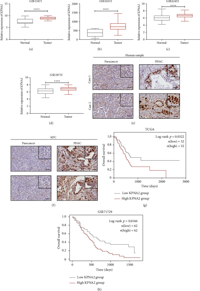
Pancreatic ductal adenocarcinoma (PDAC) is one of the deadliest malignancies and is known for its high resistance and low response to treatment. Tumor immune evasion is a major stumbling block in designing effective anticancer therapeutic strategies. Karyopherin alpha 2 (KPNA2), a member of the nuclear transporter family, is elevated in multiple human cancers and accelerates carcinogenesis. However, the specific role of KPNA2 in PDAC remains unclear. In this study, we found that expression of KPNA2 was significantly upregulated in PDAC compared to adjacent nontumor tissue and its high expression was correlated with poor survival outcome by analyzing the GEO datasets. Similar KPNA2 expression pattern was also found in both human patient samples and KPC mouse models through IHC staining. Although KPNA2 knockdown failed to impair the vitality and migration ability of PDAC cells in vitro, the in vivo tumor growth was significantly impeded and the expression of immune checkpoint ligand PD-L1 was reduced by silencing KPNA2. Furthermore, we uncovered that KPNA2 modulated the expression of PD-L1 by mediating nuclear translocation of STAT3. Collectively, our data suggested that KPNA2 has the potential to serve as a promising biomarker for diagnosis in PDAC.


Conflict of interest statement

The authors declare that they have no conflict of interest.

Figures

Figure 1
Figure 1
Expression pattern of KPNA2 in PDAC. (a–d) Expression analysis of KPNA2 in tumors and corresponding adjacent nontumor tissues using four independent GEO datasets (GSE15471, GSE16515, GSE62452, and GSE28735) (Student's t-test, ∗∗∗∗P < 0.0001). (e) IHC staining of KPNA2 expression in PDAC tumor (scale bar: 100 μm). (f) IHC staining of KPNA2 expression in the engineered mouse model of KPC (scale bar: 100 μm). (g) Kaplan-Meier overall survival (OS) curves in the TCGA dataset of PDAC patients according to the mRNA expression of KPNA2, the lower quartile value of expression was utilized as a cut-off (log-rank test, P = 0.0322). (h) Kaplan-Meier OS curve for the MPC1 expression in the GSE71729 dataset (log-rank test, P = 0.0166).
Figure 2
Figure 2
KPNA2 knockdown inhibited the growth of PDAC cells in vivo rather than in vitro. (a, b) KPNA2 silencing by sh-RNA-KPNA2 in MIA PaCa-2 and PANC-1 cells. (c) CCK8 assays of the control group and KPNA2 knockdown group of MIA PaCa-2 and PANC-1 cell lines. (d) Wound healing assays of the control group and KPNA2 knockdown group of MIA PaCa-2 and PANC-1 cell lines. (e) KPNA2 silencing by sh-RNA-KPNA2 in KPC1199-luc cell. (f) Bioluminescence imaging and luminescence intensity of orthotopic tumor growth. (g) Pancreas of C57BL/6N mice orthotopically transplanted stable KPNA2-knockdown KPC1199-luc cells and control cells. (h) Representative images of H&E staining of orthotopic PDAC model mice at 21 days.
Figure 3
Figure 3
Increased KPNA2 promoted PD-L1 expression and thereby contributed to tumor immune evasion. (a) Gene-immune analysis of KPNA2 in PDAC conducted on Sanger box. (b) The correlation between expression levels of KPNA2 and PD-L1 in PDAC. (c) Relative mRNA expression level of PD-L1 target genes in MIA PaCa-2 and PANC-1 cells with sh-KPNA2 or control vector. (d, e) Protein level of PD-L1 on the surface of MIA PaCa-2 and PANC-1 cells with sh-KPNA2 or control vector. (f, g) Relative mRNA expression level and protein level of PD-L1 in KPC1199-luc cells with sh-KPNA2 or control vector. (h) Protein level of PD-L1 on the surface of tumor cells derived from orthotopic PDAC model mice orthotopically transplanted KPC1199-luc cells with sh-KPNA2 or control vector. (i) CD8 infiltration and granzyme B expression in mouse tumor tissue detected by IHC (scale bar: 100 μm).
Figure 4
Figure 4
KPNA2 maintained the expression of PD-L1 by mediating nuclear translocation of STAT3. (a) GSEA analysis of KPNA2 expression in PDAC using the TCGA dataset. NES: normalized enrichment score. (b) Coimmunofluorescence of KPNA2 and STAT3 in MIA PaCa-2 and PANC-1 cells (scale bar: 10 μm). (c, d) KPNA2 knockdown could inhibit the nuclear translocation of STAT3 in MIA PaCa-2 and PANC-1 cells (scale bar: 10 μm). (e, f) The expression of nuclear p-STAT3 was detected in control and KPNA2 knockdown PDAC cells. Lamin B was used as the loading control of nuclear protein. (g) IHC staining of p-STAT3 in mouse tumor lesions inoculated with KPC1199-luc cell treatment with sh-KPNA2 or control vector (scale bar: 100 μm). (h, i) Relative mRNA expression level and protein level of PD-L1 in PDAC cells treated with 2.5 μM stattic, STAT3 inhibitor. (j) Proposed model for nuclear transporter KPNA2 promotes the PDAC progress.

Similar articles

Cited by

References

    1. Siegel R. L., Miller K. D., Jemal A. Cancer statistics, 2020. CA: a Cancer Journal for Clinicians. 2020;70(1):7–30. doi: 10.3322/caac.21590. - DOI - PubMed
    1. Ren B., Cui M., Yang G., et al. Tumor microenvironment participates in metastasis of pancreatic cancer. Molecular Cancer. 2018;17(1):p. 108. doi: 10.1186/s12943-018-0858-1. - DOI - PMC - PubMed
    1. Kim R., Emi M., Tanabe K. Cancer immunoediting from immune surveillance to immune escape. Immunology. 2007;121(1):1–14. doi: 10.1111/j.1365-2567.2007.02587.x. - DOI - PMC - PubMed
    1. Galon J., Bruni D. Tumor immunology and tumor evolution: intertwined histories. Immunity. 2020;52(1):55–81. doi: 10.1016/j.immuni.2019.12.018. - DOI - PubMed
    1. Martinez-Bosch N., Vinaixa J., Navarro P. Immune evasion in pancreatic cancer: from mechanisms to therapy. Cancers. 2018;10(1):p. 6. doi: 10.3390/cancers10010006. - DOI - PMC - PubMed

MeSH terms