

Abnormal retinal capillary blood flow in autosomal dominant Alzheimer's disease
- PMID: 33728371
- PMCID: PMC7931411
- DOI: 10.1002/dad2.12162
Abnormal retinal capillary blood flow in autosomal dominant Alzheimer's disease
Abstract
Introduction: This study characterizes retinal capillary blood flow in subjects with autosomal dominant Alzheimer's disease (ADAD)-causing mutations.
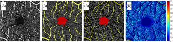
Methods: Carriers of PSEN1 or APP mutations were prospectively recruited and split into early-stage (ES) and late-stage (LS) groups. Controls were normal subjects and non-carriers from the at-risk group. Capillary blood flow was quantified using an optical coherence tomography angiography-based measure of erythrocyte flux through capillary segments. Statistical analyses were adjusted for correlation between two eyes of the same subject.
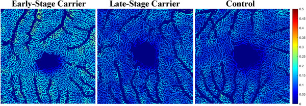
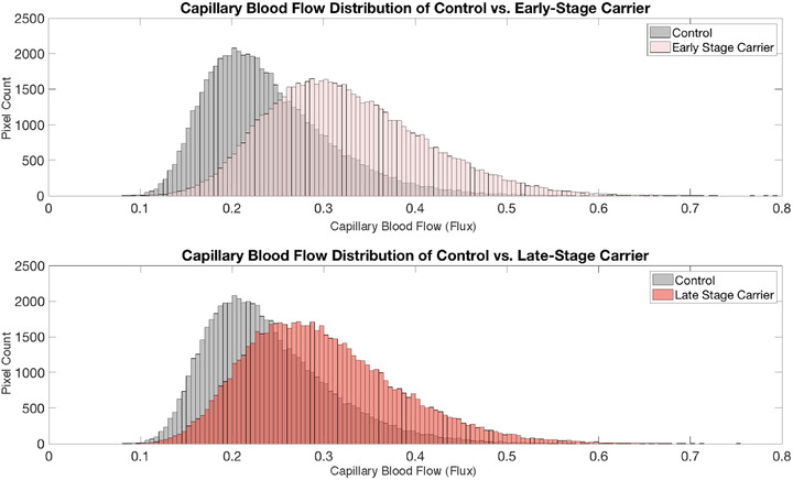
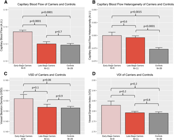
Results: ES carriers had significantly greater capillary blood flow than controls and LS carriers. ES and LS carriers had significantly greater capillary blood flow heterogeneity than controls. There was no difference between capillary blood flow of LS carriers and controls.
Discussion: ES ADAD carriers demonstrate increased retinal capillary blood flow and flow heterogeneity compared to controls. These findings support the hypothesis that increased perfusion is a pathophysiologic feature of presymptomatic stages of ADAD.
Keywords: Latino; autosomal dominant Alzheimer's disease; biomarkers; capillary blood flow; optical coherence tomography angiography; retina.
© 2021 The Authors. Alzheimer's & Dementia: Diagnosis, Assessment & Disease Monitoring published by Wiley Periodicals, LLC on behalf of Alzheimer's Association.
Conflict of interest statement
John M. Ringman is a DMC member of Renew Group Private Limited and a consultant for InnoSense. Carl Zeiss Meditec has provided grant funding, equipment, and financial support to Amir H Kashani. Ruikang K. Wang holds patents with Carl Zeiss Meditec and Kowa Inc and is a consultant for Carl Zeiss Meditec and Insight Phototonic Solutions.
Figures
References
Grants and funding
- U01 AG051218/AG/NIA NIH HHS/United States
- R01 AG054076/AG/NIA NIH HHS/United States
- P50 AG005142/AG/NIA NIH HHS/United States
- R01 EY030564/EY/NEI NIH HHS/United States
- UH2 NS100614/NS/NINDS NIH HHS/United States
- R01 AG049607/AG/NIA NIH HHS/United States
- R01 AG066524/AG/NIA NIH HHS/United States
- P30 AG066546/AG/NIA NIH HHS/United States
- R01 AG062007/AG/NIA NIH HHS/United States
- P01 AG012435/AG/NIA NIH HHS/United States
- UF1 NS100614/NS/NINDS NIH HHS/United States
- P30 AG066530/AG/NIA NIH HHS/United States
- R01 AG033193/AG/NIA NIH HHS/United States
- UH3 NS100614/NS/NINDS NIH HHS/United States
- K08 EY027006/EY/NEI NIH HHS/United States
LinkOut - more resources
Full Text Sources
Other Literature Sources