Review
. 2020 Dec;6(3):E76-E86.
doi: 10.1055/a-1347-5875. Epub 2021 Mar 12.

Contrast-Enhanced Ultrasound (CEUS) in Non-Traumatic Abdominal Emergencies

Affiliations
Review

Contrast-Enhanced Ultrasound (CEUS) in Non-Traumatic Abdominal Emergencies

Diletta Cozzi et al. Ultrasound Int Open. 2020 Dec.

Abstract

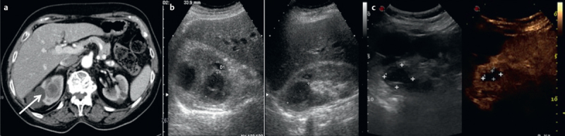
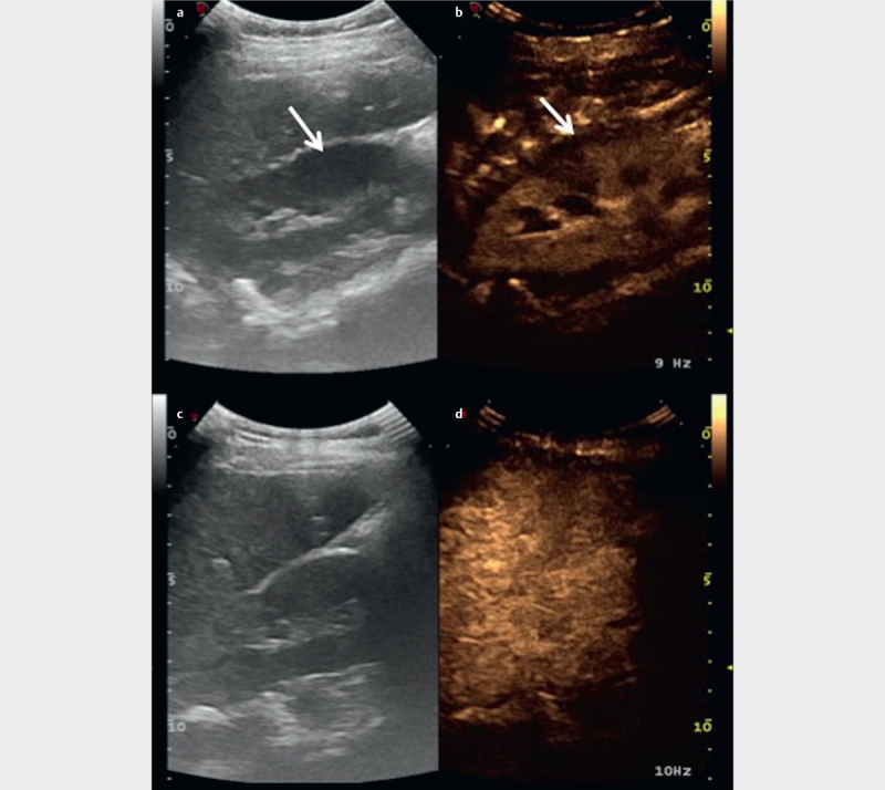
Conventional ultrasound imaging (US) is the first-line investigation in acute non-traumatic abdominal emergencies, but sometimes it needs further examinations, such as computed tomography (CT), to reach a certain diagnosis. Contrast-enhanced ultrasound (CEUS), through injection of contrast medium, may provide the radiologist with additional information that could not be investigated with baseline US. It could help reach a diagnosis and rapidly determine the proper therapy in an emergency setting. The purpose of this review is to explain and illustrate the various possibilities and limitations of CEUS in acute non-traumatic abdominal diseases, in particular acute inflammation, parenchymal infarcts, and hemorrhages.

Keywords: abdomen; acute; ultrasound; CT.


Conflict of interest statement

Conflict of Interest The authors declare that they have no conflict of interest related to the publication of this article.

Figures

Fig. 1
Fig. 1
Hemorrhagic corpus luteum. A young girl 14 y.o. with pelvic pain arrives at the emergency department and the US examination shows the presence of an enlarged left ovary compared to the contralateral. The CEUS exam shows the presence of anechoic (internal) cystic formation with an enhancing wall and an interruption of the peripheral profile (white arrow), findings compatible with rupture of the corpus luteum. It is also associated with free fluid in the pelvis.
Fig. 2
Fig. 2
Splenic pseudoaneurysm. Young male patient 18 y.o. arrives at the emergency department with upper-left quadrant pain. Baseline US (arrows in a–b ) demonstrates the presence of at least two parenchymal anechogenic lesions with peripheral vascular signal at Doppler exam. CEUS ( d–e ) and CT (C) confirm the presence of two vascular lesions within the splenic parenchyma referable to pseudoaneurysm. The diagnosis of certainty and treatment were then made with angiography and embolization of pseudoaneurysms ( f ).
Fig. 3
Fig. 3
Spontaneous HCC rupture. Case of spontaneous bleeding from an unknown liver lesion in a patient without additional comorbidities, which turned out to be hepatocellular carcinoma. CT with contrast medium ( a–b–d–e ) highlights the bleeding lesion in the left lobe, then confirmed by the CEUS examination (arrow in f ) where there is a subcapsular arterial blush indicative of bleeding in progress, which also extends into the adjacent peritoneal cavity. Angiography confirms the presence of active bleeding ( c ), embolized and no longer evident in angiographic ( h ) and CEUS controls ( g ).
Fig. 4
Fig. 4
Kidney infarct. Right cortical mesorenal infarction with triangular morphology, poorly visualized at baseline US ( a ) but without enhancement at CEUS (white arrow - b ) compared to the remaining vascularized parenchyma.
Fig. 5
Fig. 5
Acute testicular infarct. Segmental ischemia of a testicle visualized as a diffusely hypoechoic area with poor Doppler representation (white arrow in a–b–c ). The infarcted part of the testicle is not vascularized or enhanced on CEUS ( d ).
Fig. 6
Fig. 6
Acute ovarian torsion. Young girl 10 y.o. comes to the emergency department complaining of constant pain during defecation. The first US examination ( a–b ) demonstrates the presence of a coarse cystic mass within the right ovary, with slightly thickened walls with poor peripheral vascularization. CEUS ( d ) demonstrates the presence of a cystic lesion with poorly enhanced walls and with solid endoluminal parietal projections. The patient completed the diagnostic procedure with MRI (axial c , sagittal e and coronal f T2-weighted scans) which confirmed the presence of the adnexal lesion (arrow). Note the perfect agreement between the ultrasound images, CEUS, and MRI. The lesion turned out to be a cystic teratoma.
Fig. 7
Fig. 7
Acute complicated cholecystitis. On the baseline US images ( a and c) there are recognizable signs of cholecystitis with a gallbladder filled with sludge and microcalculi (arrow). CEUS highlights the evident enhancement of the gallbladder wall, which appears very thickened ( c and d ), without showing signs of complication such as an abscess or perforation.
Fig. 8
Fig. 8
Acute diverticulitis. In a , the intestinal wall is visualized an axial scan, highlighting a hypoechoic exophytic lesion suggestive of a diverticulum. After administration of US contrast medium b , the evident enhancement of the inflamed intestinal wall, in the arterial phase (arrow).
Fig. 9
Fig. 9
Abscess in diverticulitis. Pain in the left iliac fossa in a man without further comorbidity. In the clinical suspicion of an abscess, a CEUS ( a–b ) examination was performed directly: it shows a nonhomogeneous constantly anechogenic fluid collection with marked peripheral arterial enhancement, typical of an abscess. The finding was confirmed by CT ( c–d ) which also makes small air bubbles stand out better, in the context of a peri-diverticular abscess.
Fig. 10
Fig. 10
Abscess in Crohn’s disease. Similarly to Fig. 9 , this is a case of a 37-year-old male patient with Crohn’s disease with an intestinal abscess tightly attached to the abdominal wall (arrow).
Fig. 11
Fig. 11
Renal abscess. CT a and US ( b–c ) signs of a renal abscess in the upper right pole in an elderly patient (arrow). The CEUS study ( c ) confirms the presence of an abscess with an anechoic necrotic center and peripheral enhancement, as demonstrated by the CT examination. The basic US scan b does not allow complete visualization of the pathology.
Fig. 12
Fig. 12
Acute pyelonephritis. The basic US image in a highlights a blurred hypoechoic area in the right kidney. After the administration of contrast medium, the CEUS examination b confirms the presence of a triangular area with reduced enhancement compared to the remaining parenchyma: the finding refers to an area of pyelonephritis (arrows). It is also associated with perirenal free fluid collection. The images in c and d are images from a follow up CEUS study after twenty days of antibiotic therapy.

References

    1. Stoker J, Van Randen A, Laméris W et al.Imaging patients with acute abdominal pain. Radiology. 2009;253:31–46. - PubMed
    1. Choe J, Wortman J R, Micheals A et al.Beyond appendicitis: Ultrasound findings of acute bowel pathology. Emerg Radiol. 2019;26:307–317. - PubMed
    1. Van Randen A, Laméris W, Van Es H W et al.A comparison of the accuracy of ultrasound and computed tomography in common diagnoses causing acute abdominal pain. Eur Radiol. 2011;21:1535–1545. - PMC - PubMed
    1. Hunt D, Romero J. Contrast-enhanced ultrasound. Magn Reson Imaging Clin N Am. 2017;25:725–736. - PubMed
    1. Farina R, Catalano O, Stavolo C et al.Emergency radiology. Radiol Med. 2015;120:73–84. - PubMed