Review
. 2021;3(2):76-91.
doi: 10.1038/s42254-020-00267-1. Epub 2021 Jan 12.

Physics of viral dynamics

Affiliations
Review

Physics of viral dynamics

Robijn F Bruinsma et al. Nat Rev Phys. 2021.

Abstract

Viral capsids are often regarded as inert structural units, but in actuality they display fascinating dynamics during different stages of their life cycle. With the advent of single-particle approaches and high-resolution techniques, it is now possible to scrutinize viral dynamics during and after their assembly and during the subsequent development pathway into infectious viruses. In this Review, the focus is on the dynamical properties of viruses, the different physical virology techniques that are being used to study them, and the physical concepts that have been developed to describe viral dynamics.

Keywords: Biological physics; Nanoscale biophysics; Self-assembly; Supramolecular assembly; Virology.


Conflict of interest statement

Competing interestsG.J.L.W. declares financial interest in fluorescence optical tweezers and acoustic force spectroscopy approaches which are patented and licensed to LUMICKS B.V.

Figures

Fig. 1
Fig. 1. Assembly of empty capsids.
a | Phase diagram of tobacco mosaic virus (TMV) assembly. The red dot signifies cellular conditions. b | Light-scattering experiments yield the Rayleigh ratio R as a function of time for increasing capsid protein concentration (bottom to top) during assembly of human papillomavirus (HPV) virus-like particles. The mass average molecular weight of assembled particles can be determined from R and the concentration of the solution. c | High-speed atomic force microscopy snapshots of reversible capsid lattice assembly for human immunodeficiency virus (HIV) (lower panels). Scale bar, 10 nm. The top image shows a reconstruction of seven hexamers; the centre one is highlighted within a green hexagon. d | Resistive-pulse sensing technique to study hepatitis B virus (HBV) assembly, including complete T = 3 and T = 4 capsids and assembly intermediates (pre-T = 4). T, triangulation number. (Δi/i): normalized pulse amplitudes, with the baseline current i and the adjusted pulse amplitude Δi. Upper panel, integrated counts; lower panel, individual data points. Part a adapted with permission from ref.. Part b adapted with permission from ref.. Part c adapted with permission from ref.. Part d adapted with permission from ref..
Fig. 2
Fig. 2. Assembly around a genome.
a | Tobacco mosaic virus (TMV) capsid proteins (CP) assemble into A-proteins and subsequently disks. By insertion of the RNA (black thread in main image; red in inset) into a protein disk, a conformational change to a helical ‘lock-washer’ configuration occurs. The virion grows by the sequential addition of protein disks and A-proteins. b | Simulated assembly around a genome (red) following an en masse (upper) or nucleation-and-growth pathway where the genome acts as an ‘antenna’ (lower). c | Assembly traces of individual MS2 bacteriophage particles for different capsid protein concentrations, recorded by interferometric scattering microscopy. Inset: images of a single assembling particle. d | Fluorescence optical tweezers measurements of DNA packaging by synthetic capsid proteins. Progressive virus-like particle (VLP) assembly decreases the DNA end-to-end distance and pulls the beads together (left panel). Scale bar 2.5 µm. The fluorescence intensity profiles reflect the increase in bound polypeptides (right panel). e | Acoustic force spectroscopy data reveals the decrease in DNA contour length during assembly of simian virus 40 (SV40) VLPs. f | Electron micrograph (top) and corresponding schematic (bottom) of cowpea chlorotic mottle virus (CCMV) assembly around brome mosaic virus (BMV) genome. The images show (left to right): RNA without protein; decrease in size of the complex after adding capsid protein to the RNA; capsid formation after a reduction in pH. g | Time evolution of mean number of subunits 〈Nup (black circles) and radius of gyration Rg (grey circles) after mixing of CCMV capsid proteins and genome at a mass ratio ρ of 6:1. The dashed lines are decay functions for the binding time τbind (red) and the structural relaxation time τstruc (blue). Error bars indicate standard error of the mean. Part a adapted with permission from ref. and T. Splettstößer. Part b adapted with permission from ref.. Part c adapted with permission from ref.. Part d adapted with permission from ref.. Part e adapted with permission from ref.. Part f adapted with permission from ref. and ref.. Part g adapted with permission from ref..
Fig. 3
Fig. 3. Packaging signals.
A | HIV packaging efficiency of RNA containing the packaging signal Ψ, compared with RNA without Ψ (control). B | Asymmetric structure of the genome of bacteriophage MS2. C | Schematic of MS2 assembly: A-protein (AP) attaches to RNA and to capsid protein dimers (CP2) (panel Ca); additional CP2 binds and capsid formation starts (panel Cb); CP2 is not only recruited by existing RNA stem-loops (SLs) but also triggers SL formation (panel Cc); further condensation of the RNA and closed-shell formation (panel Cd); formation of a stable virion (panel Ce). D | Hamiltonian path analysis. Reconstruction of RNA inside MS2 (panel Da); the RNA shell as polyhedral cage (panel Db); 3D view of the Hamiltonian path (panel Dc); planar representation of the same Hamiltonian path with quasi-equivalent MS2 capsid subunits (panel Dd). Part A adapted with permission from ref.. Part B adapted with permission from ref.. Part C adapted with permission from ref.. Part D adapted with permission from ref..
Fig. 4
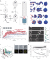
Fig. 4. Closed-shell dynamics.
a | Atomic force microscopy (AFM) image of human immunodeficiency virus (HIV) capsid with opening (rectangle) due to reverse transcription. Inset: electron microscopy image of a different particle with an opening. Scale bars, 50 nm. b | Phages HK97 (top left) and λ (bottom left) strengthen the same parts of their icosahedral shells, as visualized by these reconstructions highlighting the differences between the two particles. AFM nanoindentation curves (right panels) show the difference in mechanical response of HK97 Prohead II and Head II. Error bars indicate standard error of the mean. c | Schematic of the effect of maturation in adenovirus. d | Cowpea chlorotic mottle virus (CCMV) nanoindentation in silico, showing the first two collective excitation modes (black arrows). Pentamers are shown in blue and hexamers in red. e | Phase diagram of capsid soft modes with the dimensionless temperature β−1 as a function of the Föppl–von Kármán number γ. Vertical blue bars indicate the regions of phase coexistence of solid and molten states. Part a adapted with permission from ref.. Part b adapted with permission from refs,. Part c adapted with permission from ref.. Part d adapted with permission from ref.. Part e adapted with permission from ref..

References

    1. Baker TS, Olson NH, Fuller SD. Adding the third dimension to virus life cycles: three-dimensional reconstruction of icosahedral viruses from cryo-electron micrographs. Microbiol. Mol. Biol. Rev. 1999;63:862. doi: 10.1128/MMBR.63.4.862-922.1999. - DOI - PMC - PubMed
    1. Caspar DLD, Klug A. Physical principles in construction of regular viruses. Cold Spring Harb. Symp. Quant. Biol. 1962;27:1–24. doi: 10.1101/SQB.1962.027.001.005. - DOI - PubMed
    1. Roos WH, Bruinsma R, Wuite GJL. Physical virology. Nat. Phys. 2010;6:733–743. doi: 10.1038/nphys1797. - DOI
    1. Grunewald K, et al. Three-dimensional structure of herpes simplex virus from cryo-electron tomography. Science. 2003;302:1396–1398. doi: 10.1126/science.1090284. - DOI - PubMed
    1. Wan W, et al. Structure and assembly of the Ebola virus nucleocapsid. Nature. 2017;551:394–397. doi: 10.1038/nature24490. - DOI - PMC - PubMed

LinkOut - more resources