

Laser microgrooved vs. machined healing abutment disconnection/reconnection: a comparative clinical, radiographical and biochemical study with split-mouth design
- PMID: 33728493
- PMCID: PMC7966690
- DOI: 10.1186/s40729-021-00301-6
Laser microgrooved vs. machined healing abutment disconnection/reconnection: a comparative clinical, radiographical and biochemical study with split-mouth design
Abstract
Background: Repeated removal and replacement of healing abutments result in frequent injuries to the soft tissues.
Purpose: The purpose of this study was to evaluate the effect of disconnection/reconnection of laser microgrooved vs. machined healing and prosthetic abutments on clinical periodontal parameters, marginal bone levels, and proinflammatory cytokine levels around dental implants.
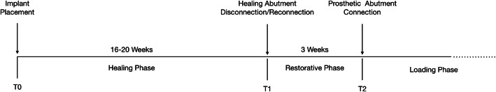
Material and methods: Twenty-four patients each received 2 implants with one-stage protocol in a split-mouth design on the same jaw. In each patient, one healing and prosthetic abutments with a laser microgrooved surface (LMS group) and one healing and prosthetic abutments with machined surface (MS group) were used. Four months following implant placement (T0), the healing abutments were disconnnected and reconnected three times to carry out the impression procedures and metal framework try-in. Four weeks later (T1), definitive prosthetic abutments were installated with screw-retained crowns. Modified plaque index (mPI), modified gingival index (mGI) bleeding on probing (BOP), and probing depth (PD) were recorded at T0 and T1. At the same time points, samples for immunological analyses were taken from the sulcus around each implant. Peri-implant crevicular fluid (PICF) samples were analyzed for interleukin-1beta (IL-1β), interleukin-6 (IL-6), and tumor necrosis factor (TNF)-α levels using the ELISA kit.
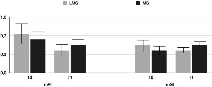
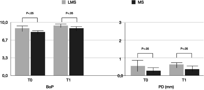
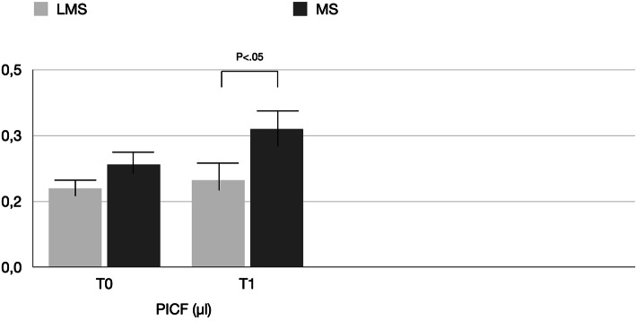
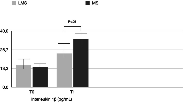
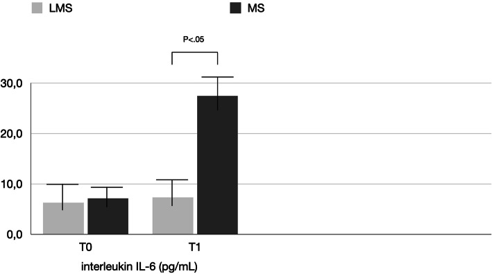
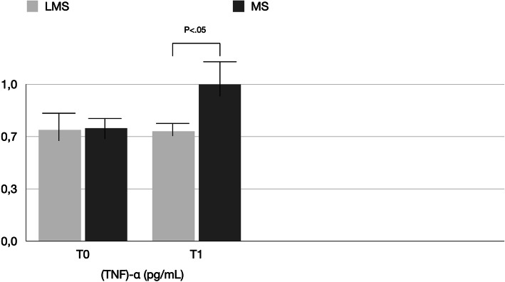
Results: At T0 and T1, mPI and mGI showed no statistical difference between the two groups, while higher PD and BoP values were noted for the MS group (P < 0.05). The mean PICF volume and mean concentrations of IL-1β, IL-6, and (TNF)-α in the LMS group were statistically less than those in the MS group (P < 0.05). In addition, comparison of IL-6 and IL-1β mean concentrations at T0 and T1 in the MS group showed a statistically significant increase (p < 0.05) over time, which was not noted for the LMS.
Conclusion: Disconnection/reconnection of healing and prosthetic abutments with a laser-microgrooved surface resulted in less inflammatory molecular response compared with conventional machined ones.
Trial registration: ClinicalTrials.gov NCT04415801 , registered 03/06/2020.
Keywords: Abutments; Cytokines; Inflammation; Peri-implant crevicular fluid.
Conflict of interest statement
Renzo Guarnieri, Gabriele Miccoli, Rodolfo Reda, Alessandro Mazzoni, Dario Di Nardo, and Luca Testarelli state that they have no competing interests.
Figures
Similar articles
-
Cross-sectional evaluation of clinical and immunological parameters at partially microgrooved vs machined abutments in humans.Int J Implant Dent. 2021 May 25;7(1):46. doi: 10.1186/s40729-021-00329-8. Int J Implant Dent. 2021. PMID: 34031775 Free PMC article.
-
Sulcus fluid volume, IL-6, and Il-1b concentrations in periodontal and peri-implant tissues comparing machined and laser-microtextured collar/abutment surfaces during 12 weeks of healing: A split-mouth RCT.Clin Oral Implants Res. 2022 Jan;33(1):94-104. doi: 10.1111/clr.13868. Epub 2021 Oct 16. Clin Oral Implants Res. 2022. PMID: 34624157 Clinical Trial.
-
Molecular Assessment of Human Peri-implant Mucosal Healing at Laser-Modified and Machined Titanium Abutments.Int J Oral Maxillofac Implants. 2018 Jul/Aug;33(4):895-904. doi: 10.11607/jomi.6367. Int J Oral Maxillofac Implants. 2018. PMID: 30025007
-
Clinical outcomes of laser microtextured implants or abutments: A systematic review.Int J Oral Implantol (Berl). 2021 May 12;14(2):141-154. Int J Oral Implantol (Berl). 2021. PMID: 34006078
-
Effect of TiO2 Abutment Coatings on Peri-Implant Soft Tissue Behavior: A Systematic Review of In Vivo Studies.Int J Dent. 2024 Mar 19;2024:9079673. doi: 10.1155/2024/9079673. eCollection 2024. Int J Dent. 2024. PMID: 38533472 Free PMC article. Review.
Cited by
-
Mechanical Behavior of Five Different Morse Taper Implants and Abutments with Different Conical Internal Connections and Angles: An In Vitro Experimental Study.J Funct Biomater. 2024 Jun 28;15(7):177. doi: 10.3390/jfb15070177. J Funct Biomater. 2024. PMID: 39057299 Free PMC article.
-
Effect of laser-microtextured abutments on peri-implant outcomes: a systematic review and meta-analysis.Clin Oral Investig. 2024 Jun 19;28(7):388. doi: 10.1007/s00784-024-05785-1. Clin Oral Investig. 2024. PMID: 38898305
-
Soft tissue integration around dental implants: A pressing priority.Biomaterials. 2026 Jan;324:123491. doi: 10.1016/j.biomaterials.2025.123491. Epub 2025 Jun 9. Biomaterials. 2026. PMID: 40505390 Review.
-
A Systematic Review of Cementation Techniques to Minimize Cement Excess in Cement-Retained Implant Restorations.Methods Protoc. 2022 Jan 17;5(1):9. doi: 10.3390/mps5010009. Methods Protoc. 2022. PMID: 35076562 Free PMC article. Review.
-
Influence of rough micro-threaded and laser micro-textured implant-neck on peri-implant tissues: A systematic review.Saudi Dent J. 2023 Sep;35(6):602-613. doi: 10.1016/j.sdentj.2023.05.025. Epub 2023 Jun 2. Saudi Dent J. 2023. PMID: 37817785 Free PMC article. Review.
References
-
- Schwarz F, Ferrari D, Herten M, Mihatovic I, Wieland M, Sager M, Becker J. Effects of surface hydrophilicity and microtopography on early stages of soft and hard tissue integration at non-submerged titanium implants: an immunohistochemical study in dogs. J Periodontol. 2007;78:2171–2184. doi: 10.1902/jop.2007.070157. - DOI - PubMed
-
- Schwarz F, Herten M, Sager M, Wieland M, Dard M, Becker J. Histological and immunohistochemical analysis of initial and early osseous integration at chemically modified and conventional SLA titanium implants: preliminary results of a pilot study in dogs. Clin Oral Implants Res. 2007;18:481–488. doi: 10.1111/j.1600-0501.2007.01341.x. - DOI - PubMed
Publication types
MeSH terms
Substances
Associated data
LinkOut - more resources
Full Text Sources
Other Literature Sources
Medical
