

Age-related changes in cerebrovascular health and their effects on neural function and cognition: A comprehensive review
- PMID: 33728712
- PMCID: PMC8244108
- DOI: 10.1111/psyp.13796
Age-related changes in cerebrovascular health and their effects on neural function and cognition: A comprehensive review
Abstract
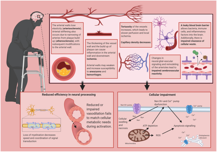
The process of aging includes changes in cellular biology that affect local interactions between cells and their environments and eventually propagate to systemic levels. In the brain, where neurons critically depend on an efficient and dynamic supply of oxygen and glucose, age-related changes in the complex interaction between the brain parenchyma and the cerebrovasculature have effects on health and functioning that negatively impact cognition and play a role in pathology. Thus, cerebrovascular health is considered one of the main mechanisms by which a healthy lifestyle, such as habitual cardiorespiratory exercise and a healthful diet, could lead to improved cognitive outcomes with aging. This review aims at detailing how the physiology of the cerebral vascular system changes with age and how these changes lead to differential trajectories of cognitive maintenance or decline. This provides a framework for generating specific mechanistic hypotheses about the efficacy of proposed interventions and lifestyle covariates that contribute to enhanced cognitive well-being. Finally, we discuss the methodological implications of age-related changes in the cerebral vasculature for human cognitive neuroscience research and propose directions for future experiments aimed at investigating age-related changes in the relationship between physiology and cognitive mechanisms.
Keywords: aging; cerebrovascular health; cerebrovascular reactivity; cognitive aging; dementia; neurovascular coupling.
© 2021 The Authors. Psychophysiology published by Wiley Periodicals LLC on behalf of Society for Psychophysiological Research.
Conflict of interest statement
The authors declare that the research was conducted in the absence of any commercial or financial relationships that could be construed as a potential conflict of interest.
Figures
Similar articles
-
The neural-vascular basis of age-related processing speed decline.Psychophysiology. 2021 Jul;58(7):e13845. doi: 10.1111/psyp.13845. Epub 2021 Jun 11. Psychophysiology. 2021. PMID: 34115388
-
Age-related alterations in the cerebrovasculature affect neurovascular coupling and BOLD fMRI responses: Insights from animal models of aging.Psychophysiology. 2021 Jul;58(7):e13718. doi: 10.1111/psyp.13718. Epub 2020 Nov 3. Psychophysiology. 2021. PMID: 33141436 Free PMC article.
-
A neural-vascular complex of age-related changes in the human brain: Anatomy, physiology, and implications for neurocognitive aging.Neurosci Biobehav Rev. 2019 Dec;107:927-944. doi: 10.1016/j.neubiorev.2019.09.005. Epub 2019 Sep 6. Neurosci Biobehav Rev. 2019. PMID: 31499083 Review.
-
Enrichment Effects on Adult Cognitive Development: Can the Functional Capacity of Older Adults Be Preserved and Enhanced?Psychol Sci Public Interest. 2008 Oct;9(1):1-65. doi: 10.1111/j.1539-6053.2009.01034.x. Epub 2008 Oct 1. Psychol Sci Public Interest. 2008. PMID: 26162004
-
Relationship between cognitive function and regulation of cerebral blood flow.J Physiol Sci. 2017 May;67(3):345-351. doi: 10.1007/s12576-017-0525-0. Epub 2017 Feb 3. J Physiol Sci. 2017. PMID: 28155036 Free PMC article. Review.
Cited by
-
Associations of Microbiota and Nutrition with Cognitive Impairment in Diseases.Nutrients. 2024 Oct 21;16(20):3570. doi: 10.3390/nu16203570. Nutrients. 2024. PMID: 39458564 Free PMC article. Review.
-
The Effect of High Fat Diet on Cerebrovascular Health and Pathology: A Species Comparative Review.Molecules. 2021 Jun 4;26(11):3406. doi: 10.3390/molecules26113406. Molecules. 2021. PMID: 34199898 Free PMC article. Review.
-
Clinicoradiological Features and Long-term Cognitive and Functional Outcome in Patients with Deep Cerebral Venous Thrombosis.Ann Indian Acad Neurol. 2024 Jan-Feb;27(1):34-39. doi: 10.4103/aian.aian_792_23. Epub 2024 Feb 1. Ann Indian Acad Neurol. 2024. PMID: 38495239 Free PMC article.
-
Effects of Aging, Estimated Fitness, and Cerebrovascular Status on White Matter Microstructural Health.Hum Brain Mapp. 2025 Apr 1;46(5):e70168. doi: 10.1002/hbm.70168. Hum Brain Mapp. 2025. PMID: 40116177 Free PMC article.
-
Functional neuroplasticity of facilitation and interference effects on inhibitory control following 3-month physical exercise in aging.Sci Rep. 2024 Feb 14;14(1):3682. doi: 10.1038/s41598-024-53974-5. Sci Rep. 2024. PMID: 38355770 Free PMC article.
References
-
- Aanerud, J. , Borghammer, P. , Chakravarty, M. M. , Vang, K. , Rodell, A. B. , Jónsdottir, K. Y. , Møller, A. , Ashkanian, M. , Vafaee, M. S. , Iversen, P. , Johannsen, P. , & Gjedde, A. (2012). Brain energy metabolism and blood flow differences in healthy aging. Journal of Cerebral Blood Flow & Metabolism, 32(7), 1177–1187. 10.1038/jcbfm.2012.18 - DOI - PMC - PubMed
-
- Abdelkarim, D. , Zhao, Y. , Turner, M. P. , Sivakolundu, D. K. , Lu, H. , & Rypma, B. (2019). A neural‐vascular complex of age‐related changes in the human brain: Anatomy, physiology, and implications for neurocognitive aging. Neuroscience & Biobehavioral Reviews, 107, 927–944. 10.1016/j.neubiorev.2019.09.005 - DOI - PubMed
-
- Ainslie, P. N. , Cotter, J. D. , George, K. P. , Lucas, S. , Murrell, C. , Shave, R. , Thomas, K. N. , Williams, M. J. A. , & Atkinson, G. (2008). Elevation in cerebral blood flow velocity with aerobic fitness throughout healthy human ageing. The Journal of Physiology, 586(Pt 16), 4005–4010. 10.1113/jphysiol.2008.158279 - DOI - PMC - PubMed
Publication types
MeSH terms
Grants and funding
LinkOut - more resources
Full Text Sources
Other Literature Sources
Medical
