. 2021 Apr;20(4):e13337.
doi: 10.1111/acel.13337. Epub 2021 Mar 16.

Mesenchymal stem cell-derived extracellular vesicles reduce senescence and extend health span in mouse models of aging

Affiliations

Mesenchymal stem cell-derived extracellular vesicles reduce senescence and extend health span in mouse models of aging

Akaitz Dorronsoro et al. Aging Cell. 2021 Apr.

Abstract

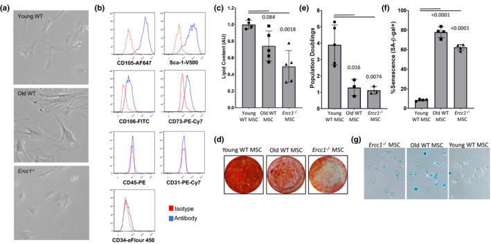
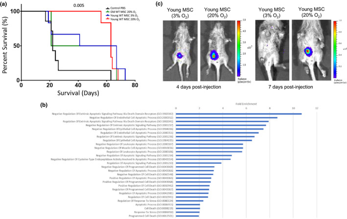
Aging drives progressive loss of the ability of tissues to recover from stress, partly through loss of somatic stem cell function and increased senescent burden. We demonstrate that bone marrow-derived mesenchymal stem cells (BM-MSCs) rapidly senescence and become dysfunctional in culture. Injection of BM-MSCs from young mice prolonged life span and health span, and conditioned media (CM) from young BM-MSCs rescued the function of aged stem cells and senescent fibroblasts. Extracellular vesicles (EVs) from young BM-MSC CM extended life span of Ercc1-/- mice similarly to injection of young BM-MSCs. Finally, treatment with EVs from MSCs generated from human ES cells reduced senescence in culture and in vivo, and improved health span. Thus, MSC EVs represent an effective and safe approach for conferring the therapeutic effects of adult stem cells, avoiding the risks of tumor development and donor cell rejection. These results demonstrate that MSC-derived EVs are highly effective senotherapeutics, slowing the progression of aging, and diseases driven by cellular senescence.

Keywords: aging; extracellular vesicles; mesenchymal stem cells; senescence; stem cells.


Conflict of interest statement

SKL is a founder of Paracrine Therapeutics, which develops EVs for therapeutic applications. PDR and LJN are co‐founders of NRTK Biosciences, which is developing approaches to reduce the senescent cell burden.

Figures

FIGURE 1
FIGURE 1
Murine bone marrow‐derived mesenchymal stem cells from naturally aged and mouse models of accelerated aging are dysfunctional. (a) Bone marrow‐derived MSCs (BM‐MSCs) from Ercc1 −/− mice are morphologically similar to BM‐MSCs from young mice and (b) share similar phenotypic membrane markers. (c) BM‐MSCs from naturally aged and Ercc1 −/− mice show impaired adipogenic potential. MSCs from different mouse models were differentiated to adipocytes using adipogenic media for 21 days. The lipid content was quantified using Nile Red stain and the values normalized to young WT mouse values. Bar graph shows mean ±SD from 3 independent experiments. Significance was determined by one‐way ANOVA (p = 0.0024, F(2,11) = 11.01) with Tukey's multiple comparisons test; p value for specific comparisons shown in figure. (d) BM‐MSCs from naturally aged and Ercc1 −/− mice have impaired osteogenic potential. MSC from the different mouse models was differentiated to osteocytes using osteogenic media for 21 days. Calcium matrix was stained using Alizarin Red S. Photographs show a differentiation representative of 3 independent experiments. (e) Proliferative potential of old WT and Ercc1 −/− BM‐MSCs is reduced compared to WT BM‐MSC controls. Significance was determined by one‐way ANOVA (p = 0.0038, F(2,8) = 12.11) with Tukey's multiple comparisons test; p values for specific comparisons shown in figure. (f) Quantitation of SA‐β‐gal staining, and representative brightfield (X‐gal) microscopy (g) of BM‐MSCs from naturally aged and Ercc1 −/− mice, which undergo senescence earlier than BM‐MSCs from young mice. The percentage of senescent cells was manually determined by counting the percent of cells positive for senescence‐associated β‐galactosidase at passage 5. Bar graph shows mean ±SD from 3 replicate representing 3 independent experiments. Significance was determined by one‐way ANOVA (p < 0.0001, F(2,9) = 394.3) with Tukey's multiple comparisons test; p values for specific comparisons shown in figure
FIGURE 2
FIGURE 2
Intraperitoneal injection of young BM‐MSCs extends life span of Ercc1 −/− mice. (a) Survival curves of Ercc1 −/− mice treated with young and old, stressed and unstressed BM‐MSCs. Approximately 106 BM‐MSCs from young and old mice were injected into Ercc1 −/− mice by intraperitoneal injection at postnatal day 10. The BM‐MSCs from young mice were maintained at 3% O2 or shifted to 20% O2 for 48 hours prior to injection. A minimum of 4 mice per group were monitored and compared against PBS‐injected controls. Survival was compared using the log‐rank Mantel‐Cox test; p value for specific comparison shown in figure. (b) Gene ontology (GO; molecular function) analysis of the transcriptome of BM‐MSCs from young mice cultured for 48 h at 20% O2 compared to BM‐MSCs maintained at 3% O2. Bars are all significant post‐FDR correction (p < 0.05) log2 fold‐enrichment values for molecular function categories related to cell survival. (c) BM‐MSCs from young mice transduced with pRETOX‐TIGHT‐GpNLuc were briefly stimulated with oxidative stress, injected IP and tracked using IVIS Xenogen imager system 4 days (left panel) and 7 days (right) post‐injection. Results shown are representative of mice from 3 independent experiments
FIGURE 3
FIGURE 3
Extracellular vesicles isolated from CM of young BM‐MSCs reduce cellular senescence and improve stem cell function in vitro and extend life span of progeroid Ercc1‐deficient mice. (a) Senescence was induced in cultures of BM‐MSCs from aged mice by passaging, and senescent cells were treated with conditioned media (CM) from the indicated BM‐MSC cultures. Bar graph shows normalized mean ±SD from 3 independent experiments. Significance was determined by one‐way ANOVA (p = 0.0002, F(3,55) = 7.630) with Tukey's multiple comparisons test; p values for specific comparisons shown in figure. (b) RT‐PCR analysis of p16INK4a expression in senescent young MSCs treated with conditioned media from functional non‐senescent MSCs; gene expression was normalized to GAPDH. Significance was determined by two‐tailed parametric unpaired t test (p = 0.0006, t(4) = 10.03); p value shown in figure. (c) Senescent cultures of ERCC1‐deficient MEFs induced by passage and oxidative stress at 20% O2 were treated with conditioned media from the indicated BM‐MSC cultures. Bar graph shows normalized mean ±SD from 3 independent experiments. ** p < 0.01. Significance was determined by one‐way ANOVA (p = 0.0003, F(2,7) = 32.69) with Tukey's multiple comparisons test; p values for specific comparisons shown in figure. (d) Electron micrograph of extracellular vesicles (EVs) in the conditioned media (CM) from BM‐MSCs from young mice. (e) Size distribution analysis of extracellular vesicles in the conditioned media from BM‐MSCs from young mice by NTA. (f) Analysis of the size of RNA content of EVs in the conditioned media from BM‐MSCs from young mice using an Agilent Bioanalyzer 2100. (g) Immunoblot analysis of Hsp70 and CD63 in EVs from conditioned media from BM‐MSCs from young mice. (h) Senescent MSCs from old mice were treated with CM or EVs isolated from the CM from BM‐MSCs from young mice. Bar graph shows normalized mean ±SD from 3 independent experiments. Significance was determined by one‐way ANOVA (p = 0.002, F(2,6) = 20.78) with Tukey's multiple comparisons test; p values for specific comparisons shown in figure. (i) Senescent BM‐MSCs from old mice were treated with CM, EVs isolated from the CM, or CM depleted of EVs from BM‐MSCs from young mice. Bar graph shows normalized mean ±SD from 3 independent experiments. Significance was determined by one‐way ANOVA (p < 0.0001, F(3,8) = 43.66) with Tukey's multiple comparisons test and t test; p values for specific comparisons shown in figure. (j) Senescent BM‐MSCs from old mice were treated with 2 x 10⁹ EV particles (light bar) or 1.25 x10¹⁰ EV particles (dark bar) from the conditioned from BM‐MSCs from young or old mice. Bar graph shows normalized mean ±SD from 3 independent experiments. Significance was determined by two‐way ANOVA with Tukey's multiple comparisons test; p values for specific comparisons shown in figure. Detailed ANOVA and multiple comparison results available in source data file. (k) IP injection of young MSC‐derived extracellular vesicles extends life span of Ercc1 −/− mice. Significance of survival curves was computed by log‐rank (Mantel‐Cox) test (p = 0.0389, χ2 = 4.265, df = 1); p value shown in figure. (l) miRNAs identified from RNA‐seq of EVs from young and old, low and high O2 cultured MSCs reveals miRNAs that are co‐regulated and enriched in young or old MSC EVs, and during oxidative stress. Significant (p < 0.05) fold‐change miRNAs are highlighted in yellow. Matrix of Pearson correlation of miRNAs expressed in all conditions reveals groups of miRNAs that are significantly (hatched cells formula image
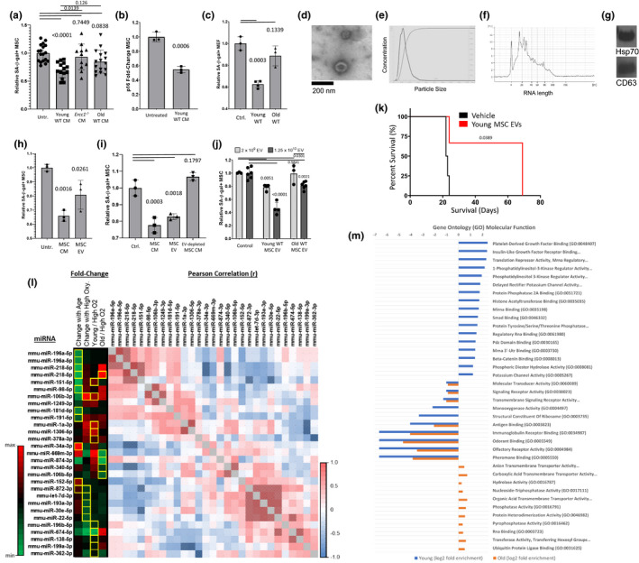
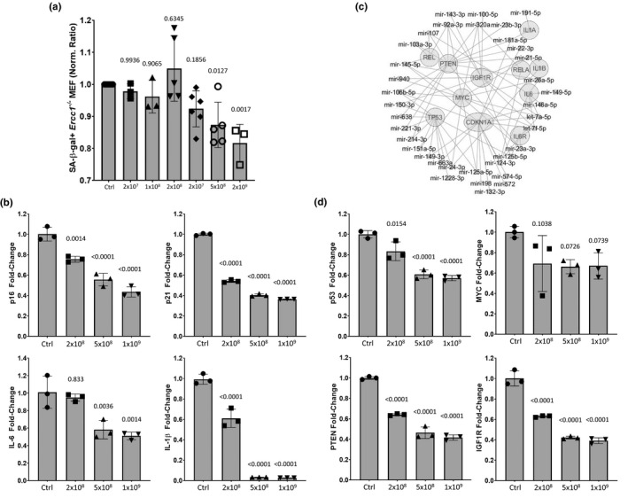
) correlated. (m) Gene ontology (GO; molecular function) of miRNA targetomes for miRNAs enriched in young and old MSC EVs; axis is log2 of fold enrichment and all categories are significant (p < 0.05) after false‐discovery rate (FDR) correction. Raw miRNA read counts, pairwise Pearson r and p values, and confidence intervals available in source data file
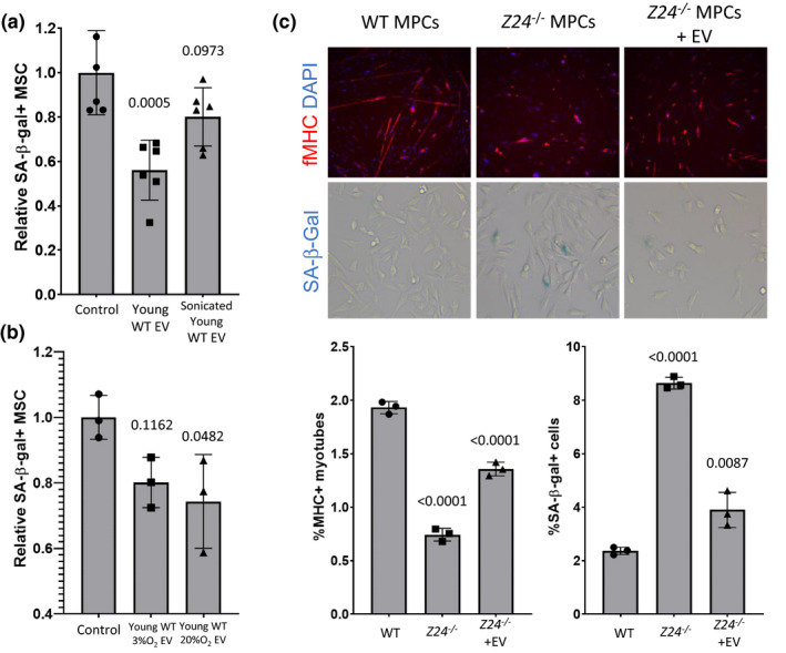
FIGURE 4
FIGURE 4
Structurally intact extracellular vesicles from young MSCs suppress senescence and restore progenitor cell function. (a) >50% senescent MSC cultures were treated with EVs from young MSCs and sonicated EVs derived from young MSC. Bar graph shows normalized mean ±SD from 3 independent experiments. Significance was determined by one‐way ANOVA (p = 0.0007, F(2,15) = 12.19) with Tukey's multiple comparisons test; p values for specific comparisons shown in figure. (b) >50% senescent MSC cultures were treated with extracellular vesicles from young MSCs and with extracellular vesicles from MSC briefly stimulated with oxidative stress. Bar graph shows normalized mean ±SD from 3 independent experiments. Significance was determined by one‐way ANOVA (p = 0.0475, F(2,6) = 5.282) with Tukey's multiple comparisons test; p values for specific comparisons shown in figure. (c) The increase in senescence in muscle progenitor cells from the Zmpste24 −/− murine model of Hutchinson Guildford progeria syndrome was reduced by treatment with EVs derived from young WT muscle‐derived progenitor cells (MDSPCs). Fluorescent microscopy images of WT and untreated and treated Zmpste24 −/− MDSPCs (upper left panel) show levels of fibrillar myosin heavy chain (fMHC, red) and nuclear DAPI staining (blue); levels of SA‐β‐gal staining for each condition are shown (blue X‐gal, lower left panel). Graphs expressing quantitation of percent myotube formation and SA‐β‐gal staining are shown (upper and lower right panel, respectively). Significance of quantitative data was determined by one‐way ANOVA: ANOVA%MHC+ (p < 0.0001, F(2,6) = 293.2) and ANOVA%SA‐β‐gal (p < 0.0001, F(2,6) = 193.9) with Tukey's multiple comparisons test; p values for specific comparisons shown in figure
FIGURE 5
FIGURE 5
Extracellular vesicles from human embryonic stem cell‐derived MSCs (AC83 EVs) reduce senescence in vitro. (a) Increasing particle numbers of AC83 EVs were added to senescent cultures of MEFs and percent senescent cells determined by C12FDG staining 72 hours post‐treatment. Significance was determined by one‐way ANOVA (p < 0.0003, F(6,24) = 6.79) with Dunnett's multiple comparisons tests; p values for specific comparisons shown in figure. (b) RT‐PCR analysis of senescent markers p16INK4a and p21Cip1 and SASP factors IL‐6 and IL‐1β measured in senescent MEFs 72 hours post‐treatment with the indicated number of AC83 EVs. Significance was determined by one‐way ANOVA: ANOVAp16 (p < 0.0001, F(3,8) = 63.34), ANOVAp21 (p < 0.0001, F(3,8) = 2277), ANOVAIL‐6 (p = 0.001, F(3,8) = 15.89) and ANOVAIL‐1β (p < 0.0001, F(3,8) = 254.3) with Dunnett's multiple comparisons test; p values for specific comparisons shown in figure. (c) Radial miRNA‐mRNA interactome demonstrates the most highly enriched miRNAs in AC83 EVs and their predicted targets. miRNAs are highly convergent upon the most centrally located targets. (d) RT‐PCR analysis of p53, PTEN, IGF‐1R, and MYC measured in senescent MEFs 24 hours post‐treatment with the indicated number of AC83 EVs. Significance was determined by one‐way ANOVA: ANOVAp53 (p < 0.0001, F(3,8) = 39.85), ANOVAMYC (p < 0.0827, F(3,8) = 3.217), ANOVAPTEN (p < 0.0001, F(3,8) = 203) and ANOVAIGF1R (p < 0.0001, F(3,8) = 145.1) with Tukey's multiple comparisons test; p values for specific comparisons shown in figure. Data shown are representative of four independent experiments with similar results
FIGURE 6
FIGURE 6
Extracellular vesicles from human embryonic stem cell‐derived MSCs (AC83 EVs) reduce markers of senescence in vivo, and suppress senescence and improve healthspan in vivo. (a) Naturally aged wild‐type mice were IP injected twice with 109 AC83 EVs. Analysis of gene expression in kidney, liver, lung, and brain by RT‐PCR revealed decreases in expression of p16INK4a and p21Cip1, IL‐6, and IL‐1β. Significance was determined by two‐tailed parametric unpaired t tests: t testp16_kidney (p < 0.0001, t(38) = 4.425), t testp16_liver (p < 0.0001, t(36) = 7.105), t testp16_lung (p = 0.91, t(69) = 0.1135), t testp16_brain (p = 0.1062, t(5) = 1.968), t testp21_kidney (p = 0.1331, t(28) = 1.547), t testp21_liver (p = 0.2895, t(38) = 1.074), t testp21_lung (p = 0.0351, t(26) = 2.223), t testp21_brain (p < 0.0001, t(26) = 5.193), t testIL‐1β_kidney (p < 0.0001, t(37) = 8.647), t testIL‐1β_liver (p < 0.0001, t(28) = 7.287), t testIL‐1β_lung (p = 0.0001, t(22) = 4.732), t testIL‐1β_brain (p < 0.0002, t(26) = 4.382), t testIL‐6_kidney (p < 0.0001, t(22) = 6.381), t testIL‐6_liver (p < 0.0001, t(19) = 4.983), t testIL‐6_lung (p = 0.3254, t(26) = 1.002), t testIL‐6_brain (p < 0.0001, t(26) = 5.920); p values shown in figure. Data shown are representative of independent experiments conducted on three cohorts of aged mice. (b) Ercc1 −/ mice expressing a p16INK4a‐luciferase reporter were given two IP injections of 109 AC83 EVs, resulting in a relative decrease in p16INKa‐mediated luciferase expression compared to control animals. Significance was determined by multiple t tests; p values shown in figure. Detailed multiple t test results available in source data file. (c) Ercc1 −/ mice given two IP injections of 109 AC83 EVs and control animals were scored for measures of health span, at 7‐ and 14‐days post‐treatment, including ataxia, body condition, dystonia, gait disorders, kyphosis and tremor. Progeroid Ercc1 −/ mice treated with AC83 EVs demonstrated consistent improvement in health span compared to control animals. Significance was determined by Šidák's multiple comparisons test; adjusted p values shown in figure. Detailed multiple comparisons test results available in source data file. Data from (b) were generated from 8, 16, 16, and 5 mice per age group, respectively. Data from (c) were generated from 4 mice per group

Similar articles

Cited by

References

    1. Abramowicz, A. , Abramowicz, A. , Widłak, P. , & Pietrowska, M. (2019). Different types of cellular stress affect the proteome composition of small extracellular vesicles: A mini review. Proteomes, 7, 23. 10.3390/proteomes7020023 - DOI - PMC - PubMed
    1. Arslan, F. , Lai, R. C. , Smeets, M. B. , Akeroyd, L. , Choo, A. , Aguor, E. N. , Timmers, L. , van Rijen, H. V. , Doevendans, P. A. , Pasterkamp, G. , Lim, S. K. , de Kleijn, D. P. (2013). Mesenchymal stem cell‐derived exosomes increase ATP levels, decrease oxidative stress and activate PI3K/Akt pathway to enhance myocardial viability and prevent adverse remodeling after myocardial ischemia/reperfusion injury. Stem Cell Research, 10(3), 301–312. 10.1016/j.scr.2013.01.002 - DOI - PubMed
    1. Baker, D. J. , Childs, B. G. , Durik, M. , Wijers, M. E. , Sieben, C. J. , Zhong, J. , A. Saltness, R. , Jeganathan, K. B. , Verzosa, G. C. , Pezeshki, A. , Khazaie, K. , Miller, J. D. , van Deursen, J. M. (2016). Naturally occurring p16(INK4a)‐positive cells shorten healthy lifespan. Nature, 530, 184–189. 10.1038/nature16932 - DOI - PMC - PubMed
    1. Baker, D. J. , Wijshake, T. , Tchkonia, T. , LeBrasseur, N. K. , Childs, B. G. , van de Sluis, B. , Kirkland, J. L. , van Deursen, J. M. (2011). Clearance of p16INK4a‐positive senescent cells delays ageing‐associated disorders. Nature, 479, 232–236. 10.1038/nature10600 - DOI - PMC - PubMed
    1. Burd, C. E. , Sorrentino, J. A. , Clark, K. S. , Darr, D. B. , Krishnamurthy, J. , Deal, A. M. , Bardeesy, N. , Castrillon, D. H. , & Beach, D. H. Sharpless, N. E. (2013). Monitoring tumorigenesis and senescence in vivo with a p16(INK4a)‐luciferase model. Cell, 152, 340–351. - PMC - PubMed

Publication types

MeSH terms