Review
. 2021 Jun 1;320(6):G919-G935.
doi: 10.1152/ajpgi.00066.2021. Epub 2021 Mar 17.

Visceral myopathy: clinical syndromes, genetics, pathophysiology, and fall of the cytoskeleton

Affiliations
Review

Visceral myopathy: clinical syndromes, genetics, pathophysiology, and fall of the cytoskeleton

Sohaib Khalid Hashmi et al. Am J Physiol Gastrointest Liver Physiol. .

Abstract

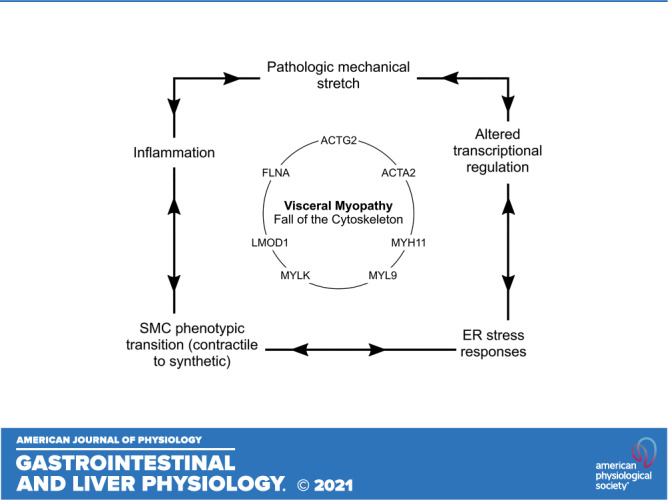
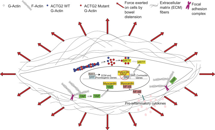
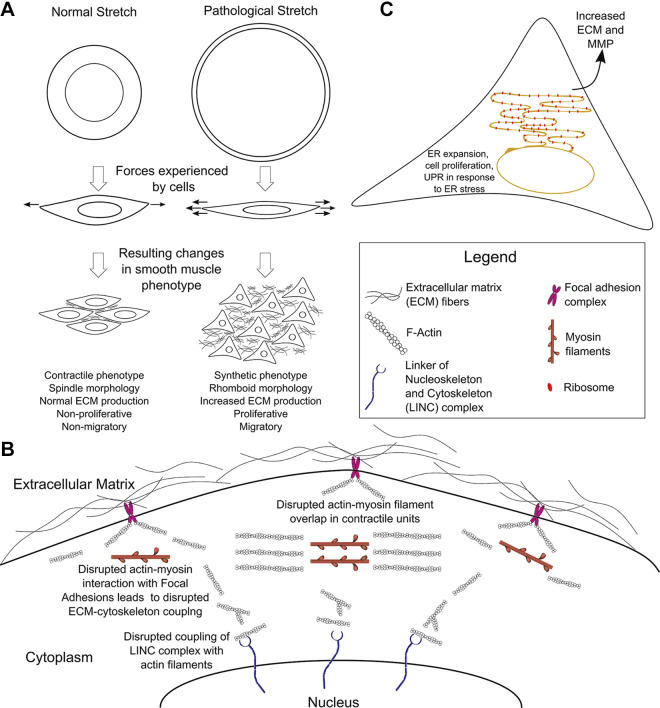
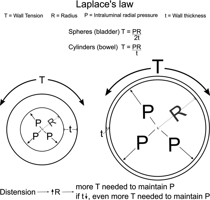
Visceral smooth muscle is a crucial component of the walls of hollow organs like the gut, bladder, and uterus. This specialized smooth muscle has unique properties that distinguish it from other muscle types and facilitate robust dilation and contraction. Visceral myopathies are diseases where severe visceral smooth muscle dysfunction prevents efficient movement of air and nutrients through the bowel, impairs bladder emptying, and affects normal uterine contraction and relaxation, particularly during pregnancy. Disease severity exists along a spectrum. The most debilitating defects cause highly dysfunctional bowel, reduced intrauterine colon growth (microcolon), and bladder-emptying defects requiring catheterization, a condition called megacystis-microcolon-intestinal hypoperistalsis syndrome (MMIHS). People with MMIHS often die early in childhood. When the bowel is the main organ affected and microcolon is absent, the condition is known as myopathic chronic intestinal pseudo-obstruction (CIPO). Visceral myopathies like MMIHS and myopathic CIPO are most commonly caused by mutations in contractile apparatus cytoskeletal proteins. Here, we review visceral myopathy-causing mutations and normal functions of these disease-associated proteins. We propose molecular, cellular, and tissue-level models that may explain clinical and histopathological features of visceral myopathy and hope these observations prompt new mechanistic studies.

Keywords: CIPO; MMIHS; cytoskeleton; visceral myopathy.


Conflict of interest statement

R. O. Heuckeroth is a consultant for BlueRock Therapeutics and served on a Scientific Advisory Panel for Takeda. None of the other authors has any conflicts of interest, financial or otherwise, to disclose.

Figures

None
Graphical abstract
Figure 1.
Figure 1.
Effects of actin gamma 2 (ACTG2) mutations and inflammation on transcriptional regulation in visceral myopathy. We propose that ACTG2 variants polymerize less efficiently than wild-type (WT) ACTG2, resulting in increased monomeric globular actin (G-actin). The increased G-actin may sequester myocardin-related transcription factors (MRTFs) in the cytoplasm, downregulating smooth muscle cell (SMC) contractile gene expression. With less MRTF in the nucleus, serum response factor (SRF) is free to bind with ELK-1, leading to increased expression of extracellular matrix (ECM) and promitogenic genes. Increased ECM deposition leads to a stiffer SMC microenvironment. This increase in stiffness along with pathologic levels of stretch due to abnormal bowel or bladder distension may increase tension on focal adhesions and release Yes-associated protein (YAP), allowing YAP to translocate to the nucleus. Nuclear YAP inhibits the interaction of myocardin with SRF, further downregulating the SMC contractile phenotype. Finally, NF-κB activation downstream of proinflammatory cytokines may further inhibit SMC contractile gene expression by NF-κB-myocardin binding. F-actin, filamentous actin.
Figure 2.
Figure 2.
Effects of mechanical stretch and endoplasmic reticulum (ER) stress responses. A: pathological stretch induces a phenotypic transition from contractile to synthetic (proliferative) phenotype in smooth muscle cells (SMCs). B: mutations that impair the actin cytoskeleton reduce force generation and inhibit development of passive tension and force transmission through the extracellular matrix (ECM). Mechanical signals are transmitted from the cell surface, through the cytoskeleton and contractile apparatus via the Linker of Nucleoskeleton and Cytoskeleton (LINC) complex to the genome, thereby affecting gene expression. If this force transmission is impaired by mutations affecting cytoskeletal filaments, physiological SMC gene expression in response to extracellular forces may be disrupted. C: endoplasmic reticulum (ER) is greatly expanded in the SMC synthetic phenotype to increase production of extracellular matrix (ECM) proteins and matrix metalloproteases (MMPs). If the cell cannot meet the increased protein production demand, an unfolded protein response (UPR) may be triggered as part of the ER stress response. This figure uses icons derived from the Reactome Icon Library by CSHL, OICR and EBI. F-actin, filamentous actin.
Figure 3.
Figure 3.
The physics of visceral myopathy. Megacystis (massive bladder dilation) and pathological bowel dilation (in chronic intestinal pseudo-obstruction, CIPO) can be modeled using Laplace’s law.
Figure 4.
Figure 4.
Hypothetical therapeutic approaches for visceral myopathies. Rapamycin and Lovastatin may increase the expression of smooth muscle cell (SMC) contractile genes, enhancing SMC force generation. Antifibrotics may make the extracellular matrix (ECM) more pliable and reduce the stiffness of the SMC microenvironment. Gene targeting approaches can be considered to silence mutations or correct them, where feasible. This figure uses icons derived from the IGI Glossary Icon Collection by Christine Liu of Two Photon Art for the Innovative Genomics Institute, and the Reactome Icon Library by CSHL, OICR and EBI.

References

    1. Thapar N, Saliakellis E, Benninga MA, Borrelli O, Curry J, Faure C, De GR, Gupte G, Knowles CH, Staiano A, Vandenplas Y, Di Lorenzo C. . Paediatric intestinal pseudo-obstruction: evidence and consensus-based recommendations from an ESPGHAN-Led Expert Group. J Pediatr Gastroenterol Nutr 66: 991–1019, 2018. doi:10.1097/mpg.0000000000001982. - DOI - PubMed
    1. Klar J, Raykova D, Gustafson E, Tóthová I, Ameur A, Wanders A, Dahl N. Phenotypic expansion of visceral myopathy associated with ACTG2 tandem base substitution. Eur J Hum Genet 23: 1679–1683, 2015. doi:10.1038/ejhg.2015.49. - DOI - PMC - PubMed
    1. Sipponen T, Karikoski R, Nuutinen H, Markkola A, Kaitila I. Three-generation familial visceral myopathy with α-actin positive inclusion bodies in intestinal smooth muscle. J Clin Gastroenterol 43: 437–443, 2009. doi:10.1097/MCG.0b013e31817d3f84. - DOI - PubMed
    1. Vargas JH, Sachs P, Ament ME. Chronic intestinal pseudo-obstruction syndrome in pediatrics. Results of a national survey by members of the North American Society of Pediatric Gastroenterology and Nutrition. J Pediatr Gastroenterol Nutr 7: 323–332, 1988. doi:10.1097/00005176-198805000-00003. - DOI - PubMed
    1. Iida H, Ohkubo H, Inamori M, Nakajima A, Sato H. Epidemiology and clinical experience of chronic intestinal pseudo-obstruction in Japan: a nationwide epidemiologic survey. J Epidemiol 23: 288–294, 2013. doi:10.2188/jea.je20120173. - DOI - PMC - PubMed

Publication types

MeSH terms

LinkOut - more resources