

Sinus computed tomography findings in patients with COVID-19
- PMID: 33729288
- PMCID: PMC7942839
- DOI: 10.31744/einstein_journal/2021AO6255
Sinus computed tomography findings in patients with COVID-19
Abstract
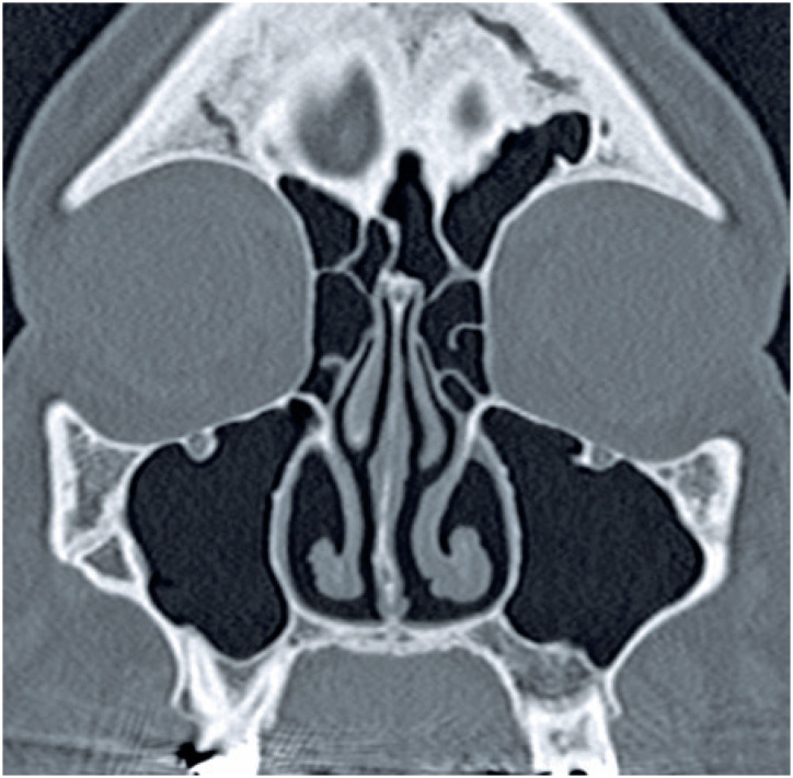
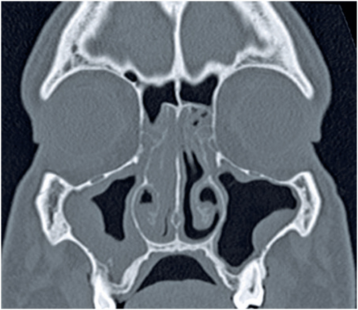
Objective: To analyze computed tomography scans of paranasal sinuses of a series of patients with coronavirus disease 2019, and correlate the findings with the disease.
Methods: Computed tomography scans of 95 adult patients who underwent a polymerase chain reaction test for severe acute respiratory syndrome coronavirus 2 were analyzed. Clinical data were obtained from patients' records and telephone calls. Paranasal sinus opacification was graded and compared according to severe acute respiratory syndrome coronavirus 2 positivity.
Results: Of the patients 28 (29.5%) tested positive for severe acute respiratory syndrome coronavirus 2 (median age 52 [range 26-95] years) and 67 were negative (median age 50 [range 18-95] years). Mucosal thickening was present in 97.4% of maxillary sinuses, 80% of anterior ethmoid air cells, 75.3% of posterior ethmoid air cells, 74.7% of frontal sinuses, and 66.3% of sphenoid sinuses. Minimal or mild mucosal thickening (score 1)and normally aerated sinuses (score 0) corresponded to 71.4% and 21.3% of all paranasal sinuses, respectively. The mean score of each paranasal sinus among severe acute respiratory syndrome coronavirus 2 positive and negative patients was 0.85±0.27 and 0.87±0.38, respectively (p=0.74). Median paranasal sinus opacification score among severe acute respiratory syndrome coronavirus 2 positive patients was 9 (interquartile range 8-10) compared to 9 (interquartile range 5-10) in negative patients (p=0.89). There was no difference in mean score adjusted for age and sex. Nasal congestion was more frequent in severe acute respiratory syndrome coronavirus 2 positive than negative patients (p=0.05).
Conclusion: Severe acute respiratory syndrome coronavirus 2 infection was associated with patient recall of nasal congestion, but showed no correlation with opacification of paranasal sinuses.
Objetivo:: Analisar imagens de tomografia computadorizada de seios paranasais de pacientes com a doença por coronavírus 2019, e correlacionar os achados com a doença.
Métodos:: Foram analisadas imagens de tomografia computadorizada de 95 pacientes submetidos a teste de reação em cadeia da polimerase para coronavírus 2 da síndrome respiratória aguda grave. Os dados clínicos foram obtidos por meio dos prontuários dos pacientes e de ligações telefônicas. A opacificação dos seios paranasais foi graduada e comparada entre pacientes positivos e negativos para coronavírus 2 da síndrome respiratória aguda grave.
Resultados:: Vinte e oito (29,5%) dos pacientes tiveram resultado positivo para coronavírus 2 da síndrome respiratória aguda grave (idade mediana de 52 [26-95] anos) e 67, resultado negativo (idade mediana de 50 [18-95] anos). O espessamento mucoso estava presente em 97,4% dos seios maxilares, 80% das células etmoidais anteriores, 75,3% das células etmoidais posteriores, 74,7% dos seios frontais e em 66,3% dos seios esfenoidais. Mínimo ou discreto espessamento mucoso (pontuação 1) e seios com aeração normal (pontuação 0) corresponderam a 71,4% e 21,3% de todos os seios paranasais, respectivamente. A nota média de cada seio paranasal entre pacientes positivos e negativos para coronavírus 2 da síndrome respiratória aguda grave foi de 0,85±0,27 e 0,87±0,38, respectivamente (p=0,74). A nota mediana de opacificação dos seios paranasais entre pacientes positivos para coronavírus 2 da síndrome respiratória aguda grave foi de 9 (intervalo interquartil de 8 a 10), comparada a 9 (intervalo interquartil de 5 a 10) em pacientes negativos (p=0,89). Não houve diferença na nota média ajustada para idade e sexo. A congestão nasal foi mais frequente em pacientes positivos para coronavírus 2 da síndrome respiratória aguda grave que naqueles com resultados negativos (p=0,05).
Conclusão:: A infecção pelo coronavírus 2 da síndrome respiratória aguda grave apresentou associação com congestão nasal, mas não mostrou correlação com espessamento mucoso dos seios paranasais.
Conflict of interest statement
none.
Figures


References
-
- 3. Guan WJ, Ni ZY, Hu Y, Liang WH, Ou CQ, He JX, Liu L, Shan H, Lei CL, Hui DS, Du B, Li LJ, Zeng G, Yuen KY, Chen RC, Tang CL, Wang T, Chen PY, Xiang J, Li SY, Wang JL, Liang ZJ, Peng YX, Wei L, Liu Y, Hu YH, Peng P, Wang JM, Liu JY, Chen Z, Li G, Zheng ZJ, Qiu SQ, Luo J, Ye CJ, Zhu SY, Zhong NS; China Medical Treatment Expert Group for Covid-19. Clinical characteristics of coronavirus disease 2019 in China. N Engl J Med. 2020;382(18):1708-20. - PMC - PubMed
MeSH terms
LinkOut - more resources
Full Text Sources
Other Literature Sources
Medical
