. 2021 Mar 1;62(3):25.
doi: 10.1167/iovs.62.3.25.

Choroidal and Retinal Changes After Systemic Adrenaline and Photodynamic Therapy in Non-Human Primates

Affiliations

Choroidal and Retinal Changes After Systemic Adrenaline and Photodynamic Therapy in Non-Human Primates

Kai Xiong Cheong et al. Invest Ophthalmol Vis Sci. .

Abstract

Purpose: To determine the tomographic, angiographic, and histologic changes in the choroid and retina of cynomolgus monkeys after systemic adrenaline and verteporfin photodynamic therapy (vPDT).

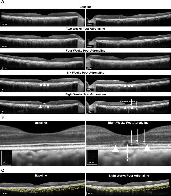
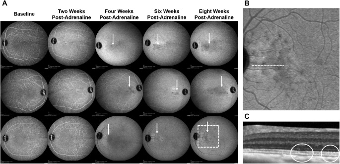
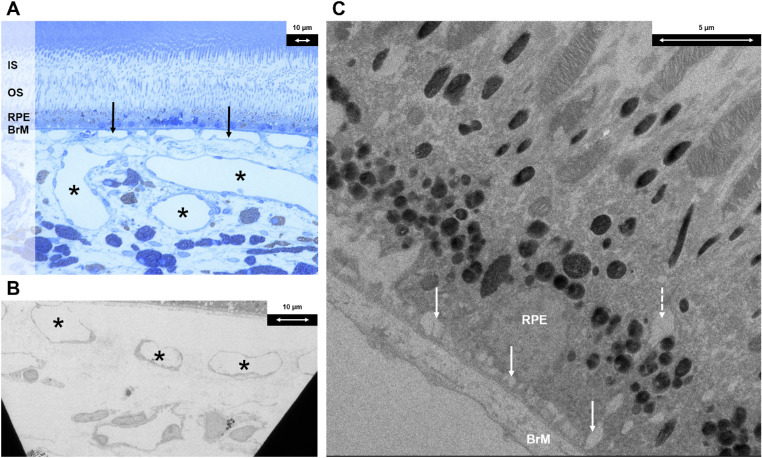
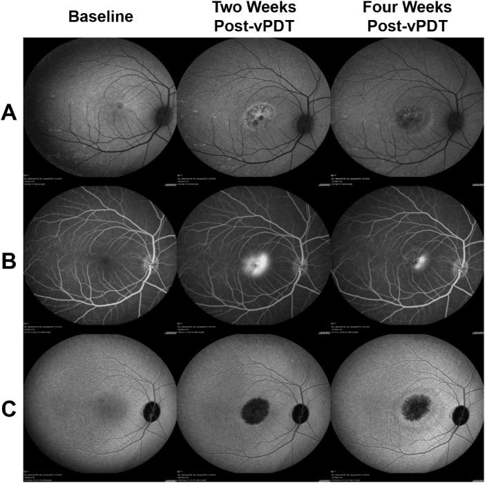
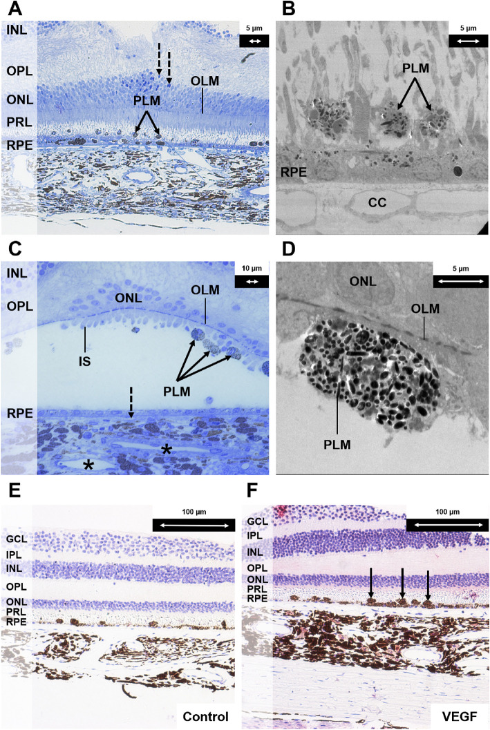
Methods: Six cynomolgus monkeys (12 eyes) were treated with vPDT only (n = 2), adrenaline only for eight weeks (n = 2), adrenaline for eight weeks with vPDT at week 4 (n = 4), and adrenaline for 12 weeks and vPDT at week 8 (n = 4). Spectral-domain optical coherence tomography, angiography, and autofluorescence were performed at baseline and every 14 days thereafter until 28 days after adrenaline therapy or vPDT. Choroid parameters included choroidal thickness (CT) changes and structural changes using semiautomated image binarization. Histology with light and electron microscopy was performed.

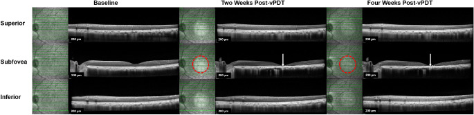
Results: Adrenaline resulted in subfoveal CT increase at week 4 compared with baseline (3.4%, P = 0.010), with further increase at week 8 (3.9%, P = 0.007). This correlated with choroidal luminal area increase (16.0% at week 8 compared with baseline, P = 0.030). Outer retinal changes included subretinal fluid, ellipsoid zone (EZ) disruption, photoreceptor elongation, and sub/intraretinal bright dots. Hypocyanescent spots surrounded by leakage was observed. Histology showed dilated choroidal vessels, intracytoplasmic vacuoles, and retinal pigment epithelium (RPE) enlarged basal infoldings. The vPDT decreased subfoveal CT at four weeks after vPDT (-7.5%, P = 0.007). This correlated with choroidal stromal area decrease (-18.0%, P < 0.010). Within the treatment spot, there was outer retinal atrophy, EZ disruption, irregular RPE thickening, intense hypoautofluorescence, hyperfluorescence, and hypocyanescence. On histology, there were outer retina, RPE, and choroid changes.

Conclusions: Adrenaline induces choroidal vessel dilation and CT increase. The vPDT decreases CT because of a reduction in choroidal stromal component.


Conflict of interest statement

Disclosure: K.X. Cheong, None; V.A. Barathi, None; K.Y.C. Teo, None; U. Chakravarthy, None; S.B.B. Tun, None; J.M. Busoy, None; C.E.H. Ho, None; R. Agrawal, None; K. Takahashi, None; C.M.G. Cheung, None

Figures

Figure 1.
Figure 1.
SD-OCT changes after systemic administration of adrenaline (group 4). (A) Longitudinal changes from baseline to eight weeks after administration of adrenaline in two eyes. From week 6 onward, disruption of the EZ was noted (indicated with white arrowheads). At eight weeks, in comparison with the baseline, there was elongation and protrusion of photoreceptor outer segments and intraretinal and subretinal bright dots (indicated with solid white arrows). There was also shallow subretinal fluid (indicated with interrupted white arrow). The areas of interest in the left eye at baseline and at week 8 are further demarcated with interrupted white outlines and are magnified in (B). (B) Magnified images comparing the left eye at week 8 after administration of adrenaline with baseline. (C) Binarization technique applied to the choroidal stromal and luminal components on the SD-OCT B-scan of the same eye at baseline and eight weeks after administration of adrenaline.
Figure 2.
Figure 2.
ICGA changes after systemic adrenaline (group 4). (A) On ICGA, round hypocyanescent spots and surrounding leakage developed from week 4 after administration of adrenaline onward in three eyes (indicated with solid white arrows). Area of interest in the last eye at week 8 is further demarcated with an interrupted white outline and is magnified in (B). (B) Magnified image of the area of interest in (A) demonstrating round hypocyanescent spots on ICGA. (C) Magnified SD-OCT B-scan of the region indicated with an interrupted white line in (B) demonstrating an effaced EZ (outlined in solid white).
Figure 3.
Figure 3.
Histologic findings following systemic adrenaline (group 2). (A) Light microscopy section with toluidine blue stain at magnification × 40 demonstrating dilated choroidal venules (indicated with black asterisks) and choriocapillaris (indicated with solid black arrows). (B) Electron microscopy section at magnification × 440 demonstrating dilated choroidal venules (indicated with black asterisks). (C) Electron microscopy section at magnification × 1900 demonstrating an intracytoplasmic vacuole at the basal aspect of RPE cells (indicated with interrupted white arrow) and enlarged basal infoldings (indicated with solid white arrows). IS, inner segment; OS, outer segment; BrM, Bruch's membrane.
Figure 4.
Figure 4.
SD-OCT changes following vPDT (group 1). Comparison of SD-OCT changes within, superior to, and inferior to vPDT treatment zone at baseline, at two weeks, and at four weeks after vPDT. Near-infrared (NIR) images are shown adjacent to the corresponding SD-OCT B-scans to indicate the location of the B-scan in relation to the vPDT treatment zone. The boundary of the vPDT treatment zone is highlighted with an interrupted red circle in the NIR image, whereas the B scan location is indicated by the solid horizontal green line. At two and four weeks after vPDT, marked outer retinal changes were observed on SD-OCT (indicated with solid white arrows). These included atrophy of outer retinal layers, severe EZ disruption, and irregular hyperreflective RPE thickening. There were no significant changes observed on SD-OCT outside the vPDT treatment zone.
Figure 5.
Figure 5.
FA, ICGA, and FAF changes after vPDT (group 1). At two weeks after vPDT, the treatment zone showed intense hypofluoresence on (A) FAF and (C) ICGA, which was surrounded by a ring of hyperfluorescence; (B) This area appeared hyperfluorescent on FA. At four weeks after vPDT, the hypofluorescence on ICGA and FAF and the hyperfluorescence on FA are partially resolved.
Figure 6.
Figure 6.
Histologic changes after half-dose half-fluence vPDT (groups 1, 3, and 4). (A) Light microscopy section with toluidine blue stain at magnification × 20 demonstrating the inner retina and choroid close to the laser spot. Pigment-laden macrophages were observed (indicated with solid black arrows). A monolayer of regenerated RPE cells with few melanin granules was seen. These cells were flatter in shape and hypopigmented because of the damage induced by vPDT (in contrast to the RPE cells in Figure 3A after administration of systemic adrenaline). A few pyknotic nuclei were seen in the ONL (indicated with interrupted black arrows). (B) Corresponding electron microscopy section at magnification × 890 demonstrating a monolayer of depigmented regenerating RPE cells after vPDT and pigment-laden macrophages (indicated with solid black arrows). (C) Composite image of light microscopy section with toluidine blue stain at magnification × 40 of the central part of the vPDT laser spot. The outer nuclear layer was disrupted. Most of the photoreceptor cells were lost. Few residual cone cells with the inner segments were seen at the foveal center. Pigment-laden macrophages were attached to the retina (indicated with solid black arrows). The RPE layer was composed by flat cells with few melanin granules. The lumens of the choriocapillaris were narrow (indicated with interrupted black arrow), and choroidal venules had narrow lumens (indicated with black asterisks). (D) Corresponding electron microscopy section at magnification × 1900 demonstrating a subretinal pigment-laden macrophage. (E) Light microscopy section at magnification × 20 as negative control for vascular endothelial growth factor (VEGF) staining of central area of vPDT. (F) Light microscopy section with VEGF antibody stain at magnification × 20 demonstrating marked VEGF staining in the choroidal stroma. There was positive staining of the subretinal macrophages (indicated with solid black arrows). There were also many round-shaped and spindle-shaped cells in the choroidal stroma compared with the control specimen. GCL, ganglion cell layer; IPL, inner plexiform layer; INL, inner nuclear layer; OPL, outer plexiform layer; ONL, outer nuclear layer; OLM, outer limiting membrane; PRL, photoreceptor layer; CC, choriocapillaris; PLM, pigment-laden macrophage.
Figure 7.
Figure 7.
Schematic of histologic changes after vPDT. Systemic administration of adrenaline resulted in an increase in CT. This was predominantly the result of an increase in LA and, to a lesser extent, an increase in SA. After vPDT, marked decrease in CT within the vPDT treatment spot resulted mainly from a reduction in SA and a less marked reduction in LA.

References

    1. Cheung CMG, Lee WK, Koizumi H, Dansingani K, Lai TYY, Freund KB.. Pachychoroid disease. Eye (Lond). 2019; 33: 14–33. - PMC - PubMed
    1. Romano MR, Parolini B, Allegrini D, Mickalewska Z, Adelman R, Bonovas S, Bopp S, EVRS study group. An international collaborative evaluation of central serous chorioretinopathy: different therapeutic approaches and review of literature. The European Vitreoretinal Society central serous chorioretinopathy study [published online ahead of print December 6, 2019]. Acta Ophthalmol, 10.1111/aos.14319. - DOI - PubMed
    1. Nicholson B, Noble J, Forooghian F, Meyerle C.. Central serous chorioretinopathy: update on pathophysiology and treatment. Surv Ophthalmol. 2013; 58: 103–126. - PMC - PubMed
    1. Kitzmann AS, Pulido JS, Diehl NN, Hodge DO, Burke JP.. The incidence of central serous chorioretinopathy in Olmsted County, Minnesota, 1980-2002. Ophthalmology. 2008; 115: 169–173. - PubMed
    1. Spaide RF, Campeas L, Haas A, et al. .. Central serous chorioretinopathy in younger and older adults. Ophthalmology. 1996; 103: 2070–2080. - PubMed

Publication types

MeSH terms

LinkOut - more resources