. 2021 Mar 1;4(3):e211740.
doi: 10.1001/jamanetworkopen.2021.1740.

Point-of-Care Digital Cytology With Artificial Intelligence for Cervical Cancer Screening in a Resource-Limited Setting

Affiliations

Point-of-Care Digital Cytology With Artificial Intelligence for Cervical Cancer Screening in a Resource-Limited Setting

Oscar Holmström et al. JAMA Netw Open. .

Abstract

Importance: Cervical cancer is highly preventable but remains a common and deadly cancer in areas without screening programs. The creation of a diagnostic system to digitize Papanicolaou test samples and analyze them using a cloud-based deep learning system (DLS) may provide needed cervical cancer screening to resource-limited areas.

Objective: To determine whether artificial intelligence-supported digital microscopy diagnostics can be implemented in a resource-limited setting and used for analysis of Papanicolaou tests.

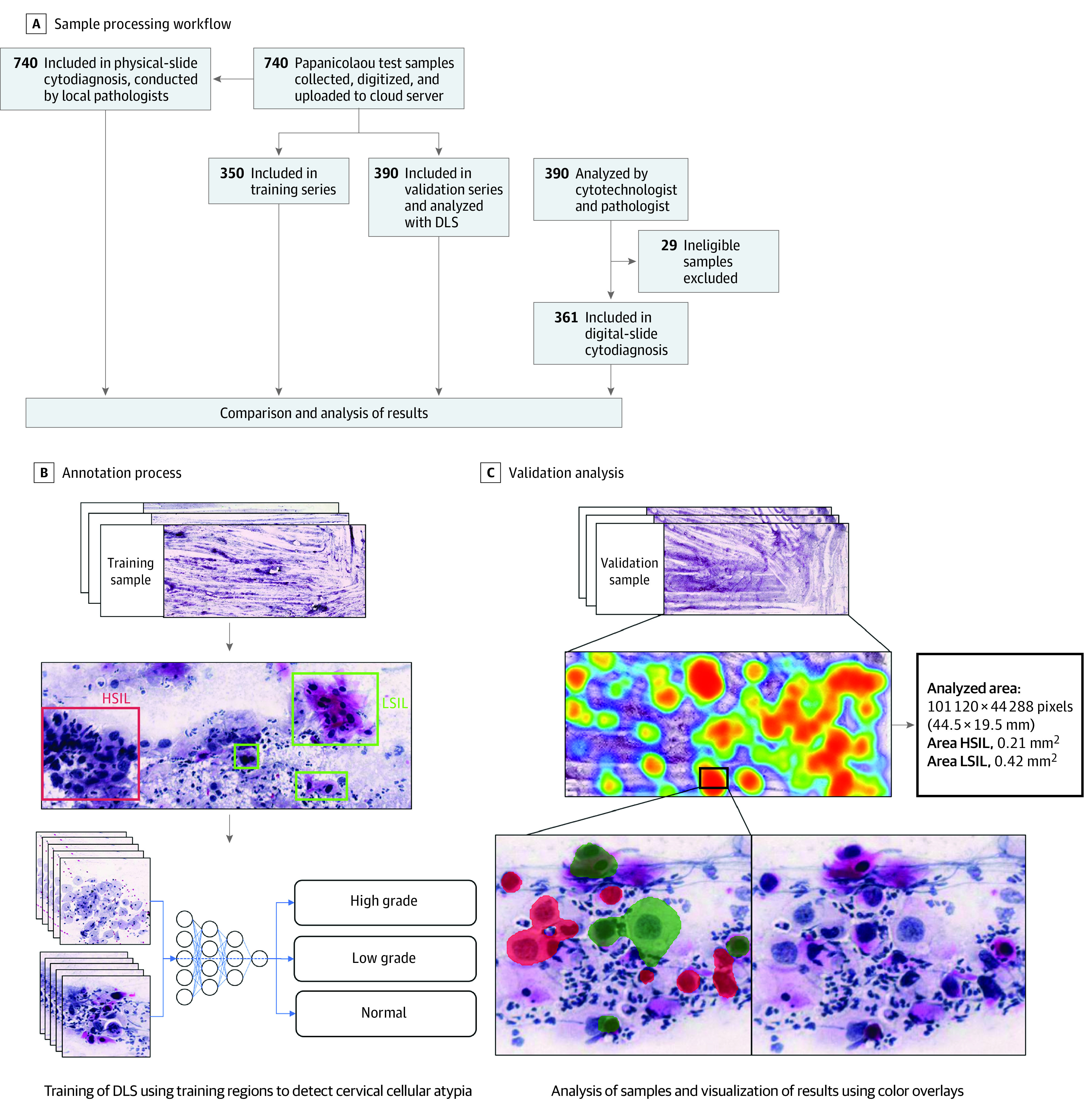
Design, setting, and participants: In this diagnostic study, cervical smears from 740 HIV-positive women aged between 18 and 64 years were collected between September 1, 2018, and September 30, 2019. The smears were digitized with a portable slide scanner, uploaded to a cloud server using mobile networks, and used to train and validate a DLS for the detection of atypical cervical cells. This single-center study was conducted at a local health care center in rural Kenya.

Exposures: Detection of squamous cell atypia in the digital samples by analysis with the DLS.

Main outcomes and measures: The accuracy of the DLS in the detection of low- and high-grade squamous intraepithelial lesions in Papanicolaou test whole-slide images.

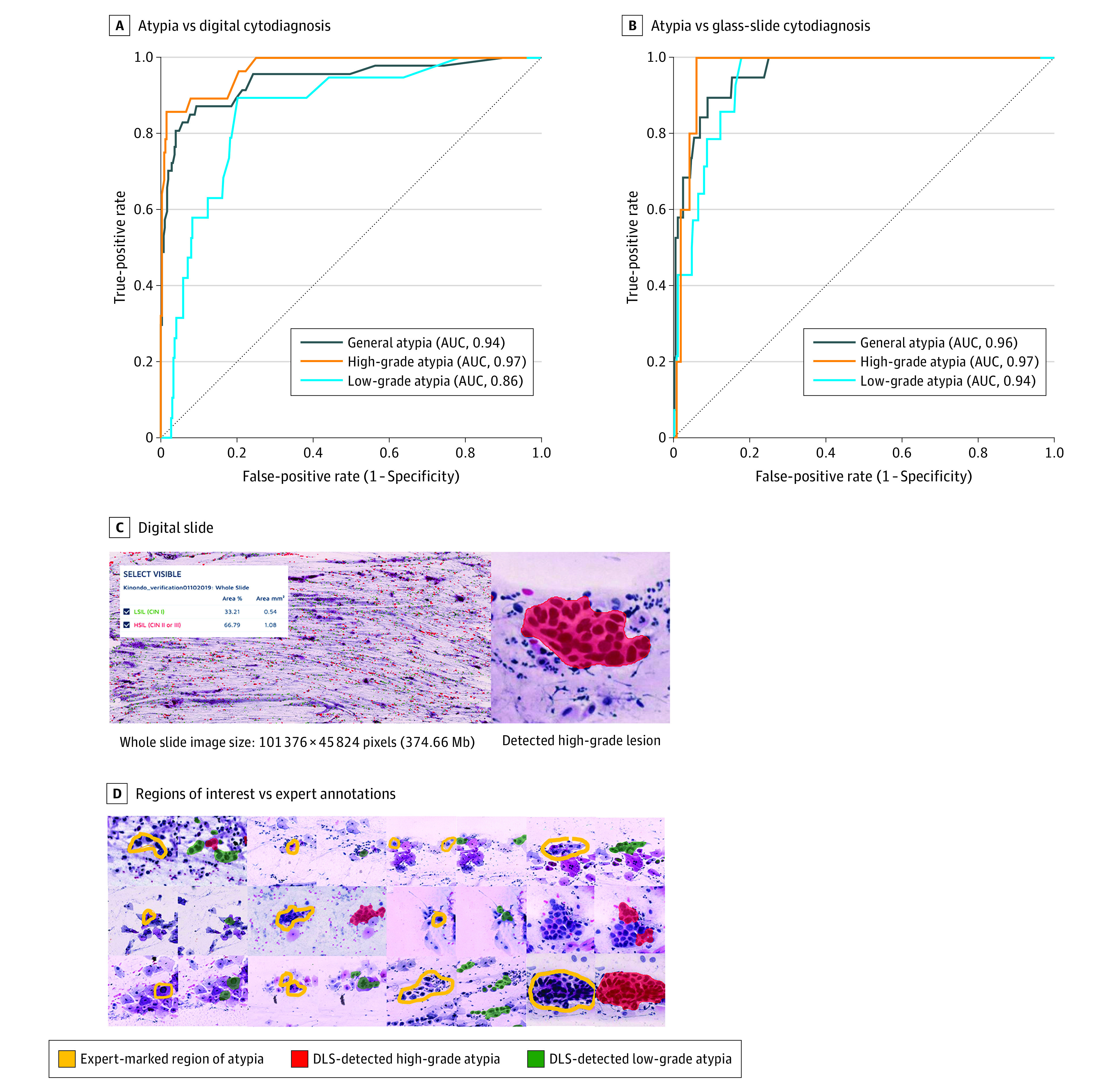
Results: Papanicolaou test results from 740 HIV-positive women (mean [SD] age, 41.8 [10.3] years) were collected. The DLS was trained using 350 whole-slide images and validated on 361 whole-slide images (average size, 100 387 × 47 560 pixels). For detection of cervical cellular atypia, sensitivities were 95.7% (95% CI, 85.5%-99.5%) and 100% (95% CI, 82.4%-100%), and specificities were 84.7% (95% CI, 80.2%-88.5%) and 78.4% (95% CI, 73.6%-82.4%), compared with the pathologist assessment of digital and physical slides, respectively. Areas under the receiver operating characteristic curve were 0.94 and 0.96, respectively. Negative predictive values were high (99%-100%), and accuracy was high, particularly for the detection of high-grade lesions. Interrater agreement was substantial compared with the pathologist assessment of digital slides (κ = 0.72) and fair compared with the assessment of glass slides (κ = 0.36). No samples that were classified as high grade by manual sample analysis had false-negative assessments by the DLS.

Conclusions and relevance: In this study, digital microscopy with artificial intelligence was implemented at a rural clinic and used to detect atypical cervical smears with a high sensitivity compared with visual sample analysis.


Conflict of interest statement

Conflict of Interest Disclosures: Dr Holmström reported receiving academic grants from Finska Läkaresällskapet, K. Albin Johanssons stiftelse, Perkléns stiftelse, Wilhelm och Elsa Stockmanns stiftelse, and Biomedicum Foundation during the 36 months before publication. Dr M. Lundin reported receiving personal fees from Aiforia Technologies Oy and serving as cofounder and co-owner of Aiforia Technologies Oy during the conduct of the study. Dr Diwan reported receiving grants from Research Council during the conduct of the study. Dr J. Lundin reported receiving personal fees from Aiforia Technologies Oy and serving as cofounder and co-owner of Aiforia Technologies Oy outside the submitted work; in addition, Dr J. Lundin reported having a patent for Mobile Microscope pending (no. WO2017037334A1; the invention is related to the use of fluorescence imaging filters combined with inexpensive plastic lenses; all rights are with the University of Helsinki) and having a patent for a slide holder for an optical microscope pending (no. WO2015185805A1; related to motorization of regular microscopes). No other disclosures were reported.

Figures

Figure 1.
Figure 1.. Practical Aspects of the Study Methodology
A, Study site location in Kenya. B, Slide processing, including staining bench and hood. C, Slide digitization equipment, including (1) laptop computer with access to the slide-management platform, (2) slide scanner, (3) mobile-network router, and (4) Papanicolaou test microscopy slide.
Figure 2.
Figure 2.. Overview of Sample Processing and Algorithm Training and Validation
A, Flowchart illustrating the sample-processing workflow, showing stages from the collection of samples to the analysis of digital images and physical slides. B, Schematic view of the annotation process used for creation of the digital-slide data for training of the deep learning system (DLS). C, Validation analysis of a digitized image of a whole slide (Papanicolaou test) with the DLS, showing calculations of areas of atypia, with locations of atypia in a heatmap of the digital slide, and identification of individual cells, with color overlays (red for high-grade atypia and green for low-grade atypia). HSIL indicates high-grade squamous intraepithelial lesions; LSIL, low-grade squamous intraepithelial lesions.
Figure 3.
Figure 3.. Detection of Atypia in Cervical Smears by Automated Deep Learning System (DLS) and by Manual Assessment
Areas under the receiver operating characteristic curves (AUCs) for the detection of general atypia, high-grade atypia, and low-grade atypia with the DLS compared with manual assessment of digital slides by a cytotechnologist and a pathologist (A) and physical slides by a local pathologist (B). Receiver operating characteristics curves were calculated for a range of operating thresholds for the DLS. C, View of a digitized sample on the cloud-based slide-management platform, with a magnified view of a detected atypical cellular cluster at 40× digital magnification. D, Examples of atypical cells marked by the experts in the digitized slides (yellow) and the corresponding regions extracted from the DLS results, with cells assessed as high-grade atypia colored in red and low-grade atypia colored in green.

Similar articles

Cited by

References

    1. Fleming KA, Naidoo M, Wilson M, et al. . An essential pathology package for low- and middle-income countries. Am J Clin Pathol. 2017;147(1):15-32. - PubMed
    1. Bogoch II, Lundin J, Lo NC, Andrews JR. Mobile phone and handheld microscopes for public health applications. Lancet Public Health. 2017;2(8):e355. doi:10.1016/S2468-2667(17)30120-2 - DOI - PubMed
    1. Arbyn M, Weiderpass E, Bruni L, et al. . Estimates of incidence and mortality of cervical cancer in 2018: a worldwide analysis. Lancet Glob Health. 2020;8(2):e191-e203. doi:10.1016/S2214-109X(19)30482-6 - DOI - PMC - PubMed
    1. Mboumba Bouassa RS, Prazuck T, Lethu T, Meye JF, Bélec L. Cervical cancer in sub-Saharan Africa: an emerging and preventable disease associated with oncogenic human papillomavirus. Med Sante Trop. 2017;27(1):16-22. doi:10.1684/mst.2017.0648 - DOI - PubMed
    1. Bosch FX, Lorincz A, Muñoz N, Meijer CJ, Shah KV. The causal relation between human papillomavirus and cervical cancer. J Clin Pathol. 2002;55(4):244-265. doi:10.1136/jcp.55.4.244 - DOI - PMC - PubMed

Publication types