Comparative Study
. 2021 Mar 17;16(3):e0247501.
doi: 10.1371/journal.pone.0247501. eCollection 2021.

Comparison of gamma and x-ray irradiation for myeloablation and establishment of normal and autoimmune syngeneic bone marrow chimeras

Affiliations
Comparative Study

Comparison of gamma and x-ray irradiation for myeloablation and establishment of normal and autoimmune syngeneic bone marrow chimeras

Thomas Rea Wittenborn et al. PLoS One. .

Abstract

Murine bone marrow (BM) chimeras are a versatile and valuable research tool in stem cell and immunology research. Engraftment of donor BM requires myeloablative conditioning of recipients. The most common method used for mice is ionizing radiation, and Cesium-137 gamma irradiators have been preferred. However, radioactive sources are being out-phased worldwide due to safety concerns, and are most commonly replaced by X-ray sources, creating a need to compare these sources regarding efficiency and potential side effects. Prior research has proven both methods capable of efficiently ablating BM cells and splenocytes in mice, but with moderate differences in resultant donor chimerism across tissues. Here, we compared Cesium-137 to 350 keV X-ray irradiation with respect to immune reconstitution, assaying complete, syngeneic BM chimeras and a mixed chimera model of autoimmune disease. Based on dose titration, we find that both gamma and X-ray irradiation can facilitate a near-complete donor chimerism. Mice subjected to 13 Gy Cesium-137 irradiation and reconstituted with syngeneic donor marrow were viable and displayed high donor chimerism, whereas X-ray irradiated mice all succumbed at 13 Gy. However, a similar degree of chimerism as that obtained following 13 Gy gamma irradiation could be achieved by 11 Gy X-ray irradiation, about 85% relative to the gamma dose. In the mixed chimera model of autoimmune disease, we found that a similar autoimmune phenotype could be achieved irrespective of irradiation source used. It is thus possible to compare data generated, regardless of the irradiation source, but every setup and application likely needs individual optimization.


Conflict of interest statement

The authors have declared that no competing interests exist.

Figures

Fig 1
Fig 1. Schematic illustration of Cesium-137 (Cs-137) gamma versus X-ray irradiation.
A) Cs-137 decays by β-emission mainly (94.6%) to the metastable nuclear isomer of Barium-137 (Ba-137), which then decays to the stable form by emitting γ-irradiation. About 5.4% of the Cs-137 will directly decay to the stable form of Ba-137. The β-irradiation does not affect the mouse since it cannot pass through the plastic container holding it, while the γ-irradiation will. B) X-rays are produced using an X-ray tube where a tungsten filament is heated and a high electrical tension is applied, rendering it a cathode which accelerates electrons (e-) at high speed towards a grounded target acting as anode. This creates a continuous spectrum of energies. The low energy photons are filtered out using a filter composed of tin (Sn), copper (Cu) and aluminum (Al). However, this also lowers the intensity of the radiation accordingly (blue curve). Be; beryllium.
Fig 2
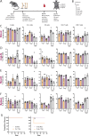
Fig 2. The irradiation of mice using Cesium-137 (Cs-137) or X-ray affects the levels of B, Gr1+, NK, CD4 T and CD8 T cells similarly.
A) Schematic illustration of experimental layout. B) Gating tree used for flow cytometry data, gating layout can be seen in S1A Fig. Percentages of immune cells in blood (C), IngLN (D), MesLN (E) and spleen (F) after Cs-137 irradiation at doses of 9 (n = 8), 11 (n = 8) and 13 (n = 6) Gy or X-ray irradiation at doses of 6.3 (n = 4), 7.7 (n = 4), 9 (n = 7) and 11 (n = 4) Gy. Naïve mice have not been exposed to any treatment. In all bar graphs, the bar indicates the mean and error bars indicate the standard error of mean. For blood measurements, Kruskal-Wallis test indicated significant differences between medians within Gr1+ cells (P = 0.01) and NK cells (P = 0.016), with significant differences for Dunn’s pairwise comparison within NK cells for 11 Gy Cs-137 vs. 7.7 Gy X-ray (P = 0.047), 11 Gy Cs-137 vs. 6.3 Gy (P = 0.0338), and 11 Gy X-ray vs. 6.3 Gy X-ray (P = 0.0495). For IngLN, Kruskall-Wallis test indicated only significantly different medians among Gr1+ cells (P = 0.035), with pairwise differences for 11 Gy X-ray vs. 6.3 Gy X-ray (P = 0.0227). For MesLN, Kruskal-Wallis test indicated significant differences between medians within Gr1+ (P = 0.0048) and CD8 T cells (P = 0.0001), with significant differences for Dunn’s pairwise comparison within Gr1+ for 11 Gy X-ray vs. 6.3 Gy X-ray (P = 0.0041) and within CD8 T cells for 13 Gy Cs-137 vs. 7.7 Gy X-ray (P = 0.0071), 9 Gy Cs-137 vs. 11 Gy X-ray (P = 0.0369), 11 Gy X-ray vs. 7.7 Gy X-ray (P = 0.0015) and 6.3 Gy X-ray (P = 0.0141). Finally, for spleen, significantly different medians were found among B (P = 0.0288), Gr1+ (P = 0.0021) and NK cells (P = 0.0046), with significant pairwise differences within B cells, 11 Gy X-ray vs. 6.3 Gy X-ray (P = 0.032), within Gr1+, 11 Gy Cs-137 vs. 6.3 Gy X-ray (P = 0.0475), 9 Gy Cs-137 vs 11 Gy X-ray (P = 0.0462), and 11 Gy X-ray vs. 6.3 Gy X-ray (P = 0.0193), and within NK cells, 11 Gy X-ray vs. 6.3 Gy X-ray (P = 0.0044). G) Survival curves for the mice after each irradiation dose. All mice receiving 13 Gy X-ray irradiation died or were euthanized as they reached a humane end-point. Statistical significance indicated for log-ranked Mantel-Cox test (***, p<0.001).
Fig 3
Fig 3. The degree of chimerism depends upon irradiation dose.
A) Schematic illustration of flow cytometry gating tree, gating layout can be seen in S1B Fig. Percentages of chimerism in blood (B), IngLN (C), MesLN (D) and spleen (E) after Cs-137 irradiation at doses 9, 11 and 13 Gy or X-ray irradiation at doses 6.3, 7.7, 9 and 11 Gy. CD45.1 positive cells come from the donor mice and CD45.2 cells are remnants of the recipients own cells. Naïve mice have not been exposed to any treatment. In all bar graphs, the bar indicates the mean and error bars indicate the standard error of mean. Based on two-way ANOVA, irradiation modality did not come out as an independent source of variation in the data for any of the tissues or cell subsets analyzed, although there was a significant interaction effect (P = or P<0.0001 in all cases).
Fig 4
Fig 4. Cesium-137 (Cs-137) or X-ray irradiation affects the levels of immune cells similarly also in an autoimmune environment.
A) Schematic illustration of experimental layout, where 1 part of the injected bone marrow came from the autoimmune mouse strain 564Igi and 2 parts came from wildtype donors. B) Gating tree for flow cytometry strategy, gating layout can be seen in S2A Fig. Percentages of immune cells in blood (C), IngLN (D), MesLN (E) and spleen (F) after irradiation at doses of 9 (n = 5) and 11 (n = 3 survivors, see G) Gy using either X-ray or Cs-137 irradiation. Naïve mice have not been exposed to any treatment. In all bar graphs, the bar indicates the mean and error bars indicate the standard error of mean. For blood measurements, Kruskal-Wallis test indicated significant differences between medians within B cells (P = 0.0403) and Gr1+ cells (P = 0.0289), but with no significant differences for Dunn’s pairwise comparison for any of the groups. For IngLN, the only cell population displaying significant differences between medians based upon Kruskal-Wallis test was the CD8 group (P = 0.0326), with Dunn’s post-test giving a significant difference between 11 Gy Cs-137 and 11 Gy X-ray (P = 0.0393). For spleen, there were significant differences between medians for NK (P = 0.001), CD4 (P = 0.0077) and CD8 (P = 0.0059) populations, but Dunn’s post-test came out significant only for 9 Gy Cs-137 vs. 11 Gy X-ray (P = 0.0134, P = 0.0372 and P = 0.0231, respectively). G) Survival curves for the mice after each irradiation dose. No statistically significant differences were noted based on log-ranked Mantel-Cox test with alpha = 0.05.
Fig 5
Fig 5. Chimerism of 564Igi mixed chimeras.
A) Schematic overview of flow cytometry gating tree, gating layout can be seen in S2B Fig. Percentages of chimerism in blood (B), IngLN (C), MesLN (D) and spleen (E) after irradiation at doses of 9 and 11 Gy using either X-ray or Cs-137 irradiation. CD45.1 positive cells derive from the donor mice and CD45.2 cells are residual recipient cells or derive from the 564Igi donor compartment. Naïve mice have not been exposed to any treatment. In all bar graphs, the bar indicates the mean and error bars indicate the standard error of mean. Based on two-way ANOVA, irradiation modality only came out as an independent source of variation in the data for the NK cell population in MesLN (P = 0.0321), but in all cases there was a significant interaction effect (P<0.0001 throughout).
Fig 6
Fig 6. Autoimmune hallmarks in the mixed bone marrow chimeras are independent of irradiation method.
Percentages of immune cells in blood (A), IngLN (B), MesLN (C) and spleen (D) after irradiation at doses of 9 and 11 Gy using either X-ray or Cs-137 irradiation, and fractional representation of CD45.1 vs. CD45.2 cells within GC B cells of IngLN, MesLN and spleen. Naïve mice have not been exposed to any treatment. Gating layout can be seen in S3 Fig. In all bar graphs, the bar indicates the mean and error bars indicate the standard error of mean. For MesLN cell frequencies, Kruskal-Wallis test indicated significant differences between medians within GC B cells (P = 0.0322), but with no significant differences for Dunn’s pairwise comparison for any of the groups. For relative representation of CD45.1 vs. CD45.2 cells, two-way ANOVA did not identify irradiation modality as an independent source of variation in the data for any of the tissues analyzed, although there was a significant interaction effect (P<0.0001 in all cases).

Similar articles

Cited by

References

    1. Riddell J, Gazit R, Garrison BS, Guo G, Saadatpour A, Mandal PK, et al.. Reprogramming committed murine blood cells to induced hematopoietic stem cells with defined factors. Cell. 2014;157: 549–564. 10.1016/j.cell.2014.04.006 - DOI - PMC - PubMed
    1. Das A, Heesters BA, Bialas A, O’Flynn J, Rifkin IR, Ochando J, et al.. Follicular Dendritic Cell Activation by TLR Ligands Promotes Autoreactive B Cell Responses. Immunity. 2017;46: 106–119. 10.1016/j.immuni.2016.12.014 - DOI - PMC - PubMed
    1. Shlomchik WD, Couzens MS, Tang CB, McNiff J, Robert ME, Liu J, et al.. Prevention of graft versus host disease by inactivation of host antigen- presenting cells. Science (80-). 1999;285: 412–415. 10.1126/science.285.5426.412 - DOI - PubMed
    1. Toubai T, Tawara I, Sun Y, Liu C, Nieves E, Evers R, et al.. Induction of acute GVHD by sex-mismatched H-Y antigens in the absence of functional radiosensitive host hematopoietic-derived antigen-presenting cells. Blood. 2012;119: 3844–3853. 10.1182/blood-2011-10-384057 - DOI - PMC - PubMed
    1. Degn SE, van der Poel CE, Firl DJ, Ayoglu B, Al Qureshah FA, Bajic G, et al.. Clonal Evolution of Autoreactive Germinal Centers. Cell. 2017;170: 913–926.e19. 10.1016/j.cell.2017.07.026 - DOI - PMC - PubMed

Publication types