. 2021 Aug;24(3):471-482.
doi: 10.1007/s10456-021-09776-8. Epub 2021 Mar 17.

Phosphorylation of pericyte FAK-Y861 affects tumour cell apoptosis and tumour blood vessel regression

Affiliations

Phosphorylation of pericyte FAK-Y861 affects tumour cell apoptosis and tumour blood vessel regression

Delphine M Lees et al. Angiogenesis. 2021 Aug.

Erratum in

Abstract

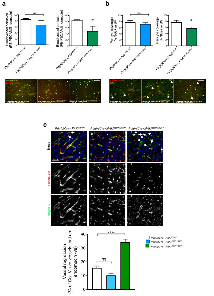
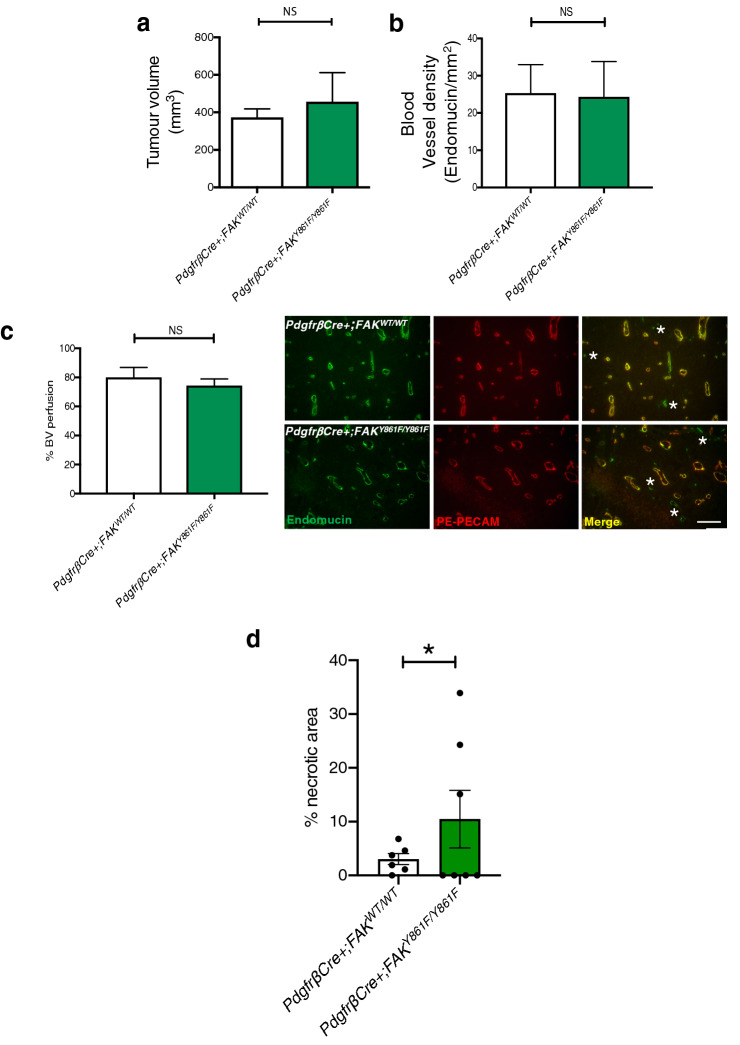
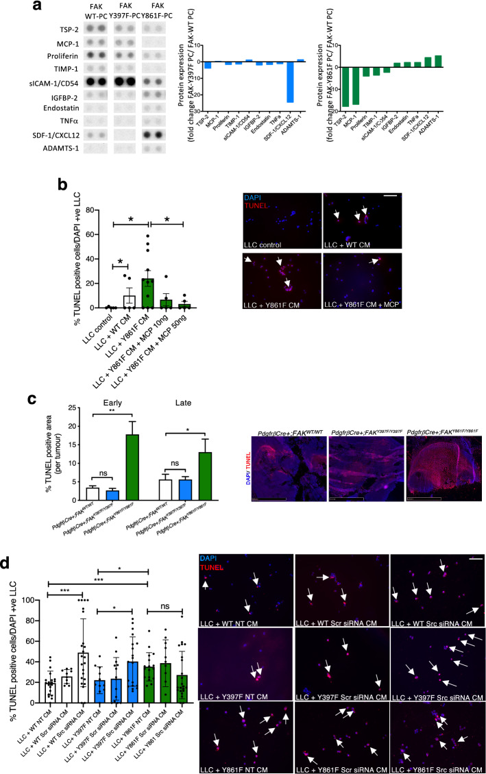
Focal adhesion kinase (FAK) is a non-receptor tyrosine kinase that is overexpressed in many cancer types and in vivo studies have shown that vascular endothelial cell FAK expression and FAK-phosphorylation at tyrosine (Y) 397, and subsequently FAK-Y861, are important in tumour angiogenesis. Pericytes also play a vital role in regulating tumour blood vessel stabilisation, but the specific involvement of pericyte FAK-Y397 and FAK-Y861 phosphorylation in tumour blood vessels is unknown. Using PdgfrβCre + ;FAKWT/WT, PdgfrβCre + ;FAKY397F/Y397F and PdgfrβCre + ;FAKY861F/Y861F mice, our data demonstrate that Lewis lung carcinoma tumour growth, tumour blood vessel density, blood vessel perfusion and pericyte coverage were affected only in late stage tumours in PdgfrβCre + ;FAKY861F/Y861F but not PdgfrβCre + ;FAKY397F/Y397F mice. Further examination indicates a dual role for pericyte FAK-Y861 phosphorylation in the regulation of tumour vessel regression and also in the control of pericyte derived signals that influence apoptosis in cancer cells. Overall this study identifies the role of pericyte FAK-Y861 in the regulation of tumour vessel regression and tumour growth control and that non-phosphorylatable FAK-Y861F in pericytes reduces tumour growth and blood vessel density.

Keywords: Angiogenesis; Cancer; Focal adhesion kinase (FAK); Pericytes.


Conflict of interest statement

KHD is a scientific advisor for Ellipses. KHD is joint applicant on Patent claims N421127GB and N417173GB. The other authors declare no conflicts of interest.

Figures

Fig. 1
Fig. 1
LLC tumour growth and angiogenesis are reduced in PdgfrβCre + ;FAKY861F/Y861F mice. a PdgfrβCre + ;FAKWT/WT, PdgfrβCre + ;FAKY397F/Y397F and PdgfrβCre + ;FAKY861F/Y861F mice were born at normal male;female ratios; b Mendelian ratios with c similar body weights. Pie chart in a represents percentage male:female ratio, in b represents % Cre + and Cre- mice born to each genotype (n = 60 mice/genotype). d Western blotting of primary pericytes isolated from PdgfrβCre + ;FAKWT/WT, PdgfrβCre + ;FAKY397F/Y397F and PdgfrβCre + ;FAKY861F/Y861F mice confirmed reduced levels of p-Y397 and pY861-FAK in FAKY397F and FAKY861F pericytes, respectively. e pY861-FAK levels are significantly reduced in Y397F pericytes, n = 2 independent lysates/genotype. Bar chart represents mean pY861-FAK levels + s.e.m. *P = 0.0155. GAPDH act as loading control. f pY416-Src levels are significantly reduced in Y397F-FAK pericytes. Blots shows pY416-Src, total Src and GAPDH loading control. g In vivo tumour growth was significantly reduced only in PdgfrβCre + ;FAKY861F/Y861F mice. Graphs represent mean tumour volume ± s.e.m.; n = 15 PdgfrβCre + ;FAKWT/WT mice, 14 PdgfrβCre + ;FAKY397F/Y397F mice and 11 PdgfrβCre + ;FAKY861F/Y861F mice. ****P < 0.0001. Two-sided Mann–Whitney U rank sum test. h Tumour blood vessel density was significantly reduced only in PdgfrβCre + ;FAKY861F/Y861F mice. Bar charts represent mean blood vessel density + s.e.m. *P = 0.0498; ns, not significant; n = 6 PdgfrβCre + ;FAKWT/WT tumours, 6 PdgfrβCre + ;FAKY397F/Y397F tumours and 5 PdgfrβCre + ;FAKY861F/Y861F tumours. Two-sided Student’s t-test. Representative endomucin stained LLC tumour sections are shown for each genotype. Scale bar, 50 μm
Fig. 2
Fig. 2
Blood vessel perfusion and pericyte coverage are reduced in PdgfrβCre + ;FAKY861F/Y861F mice. a PE-PECAM antibody perfused LLC tumours grown in PdgfrβCre + ;FAKWT/WT, PdgfrβCre + ;FAKY397F/Y397F and PdgfrβCre + ;FAKY861F/Y861F mice showed a significant reduction in functional tumour blood vessels in PdgfrβCre + ;FAKY861F/Y861F mice. Bar charts represent mean blood vessel perfusion + s.e.m. *P = 0.032; ns, not significant; n = 6 PdgfrβCre + ;FAKWT/WT tumours, 5 PdgfrβCre + ;FAKY397F/Y397F tumours and 4 PdgfrβCre + ;FAKY861F/Y861F tumours. Two-sided Student’s t-test. Representative endomucin stained and PE-PECAM antibody perfused LLC tumour sections are shown for each genotype. asterisks, non-perfused blood vessels. b Pericyte coverage of tumour blood vessels was also reduced in these mice. Bar charts represent mean pericyte coverage + s.e.m. *P = 0.03, ns, not significant; n = 6 PdgfrβCre + ;FAKWT/WT tumours, 5 PdgfrβCre + ;FAKY397F/Y397F tumours and 4 PdgfrβCre + ;FAKY861F/Y861F tumours. Two-sided Student’s t-test. Representative double stained endomucin and NG2 LLC tumour sections are shown for each genotype. Arrows, NG2 + endomucin + blood vessels; asterisks, NG2- endomucin + blood vessels. Scale bar in a and b, 50 μm. c Day 14–21 tumours from PdgfrβCre + ;FAKWT/WT, PdgfrβCre + ;FAKY397F/Y397F and PdgfrβCre + ;FAKY861F/Y861F mice were immunostained with Collagen IV and endomucin to identify empty basement membrane sheaths. Bar chart shows vessel regression (% of Coll IV + vessels that are endomucin -ve) + s.e.m.; ****P < 0.0001; ns, not significant; n = 6 PdgfrβCre + ;FAKWT/WT mice, 8 PdgfrβCre + ;FAKY397F/Y397F mice and 6 PdgfrβCre + ;FAKY861F/Y861F mice. Two-way ANOVA. Representative images show Collagen IV and endomucin stained blood vessels from tumours from all genotypes. arrows, Collagen IV + endomucin –ve blood vessels. Scale bar, 50 μm
Fig. 3
Fig. 3
Early stage tumour growth is similar between PdgfrβCre + ;FAKWT/WT and PdgfrβCre + ;FAKY861F/Y861F mice. Early (day 12): a Tumour size, b Blood vessel density, and c Blood vessel perfusion were similar between PdgfrβCre + ;FAKWT/WT and PdgfrβCre + ;FAKY861F/Y861F mice. Bar charts represent mean + s.e.m.; ns, not significant. Student’s t-test. For a, n = 8 PdgfrβCre + ;FAKWT/WT mice, 5 PdgfrβCre + ;FAKY861F/Y861F mice. For b and c n = 8 PdgfrβCre + ;FAKWT/WT tumours, 5 PdgfrβCre + ;FAKY861F/Y861F tumours. Representative images showing blood vessel perfusion in LLC tumours from PdgfrβCre + ;FAKWT/WT and PdgfrβCre + ;FAKY861F/Y861F mice. Scale bar, 50 μm. Asterisks, non-perfused blood vessels. d Tumour necrosis was increased in PdgfrβCre + ;FAK Y861F/Y861F. Bar chart shows % necrotic area in tumours from PdgfrβCre + ;FAKWT/WT and PdgfrβCre + ;FAK Y861F/Y861Fmice.*P < 0.05 Wilcoxon test; n = 6 tumours from PdgfrβCre + ;FAKWT/WT mice and 7 tumours from PdgfrβCre + ;FAK Y861F/Y861F mice
Fig. 4
Fig. 4
FAK-Y861F pericytes induce apoptosis in LLC tumour cells. a R&D proteome profiler array using lysates from FAK-WT, FAK-Y397F and FAK-Y861F pericytes. Representative dots of differentially expressed proteins are given. Bar charts show mean fold change in protein expression relative to levels in WT. N = 2 dots from 1 experiment. b Lewis lung carcinoma (LLC) cells incubated with conditioned medium (CM) from pericytes plus or minus recombinant MCP-1. Treatment with FAK-Y861F pericyte CM increased LLC apoptosis, compared with CM from FAK-WT pericytes. This effect was rescued upon treatment with MCP-1 (10 and 50 ng/ml). Bar chart represents % TUNEL-positive cells + s.e.m. Representative images show effect of CM and MCP-1 on LLC cells. Arrows, TUNEL-positive cells. *P < 0.05. One-way ANOVA. N = 5–11 fields of view/genotype. Scale bar, 500 μm. c Early and late stage tumours from PdgfrβCre + ;FAKY861F/Y861F mice had significantly larger TUNEL-positive areas than tumours from PdgfrβCre + ;FAKWT/WT and PdgfrβCre + ;FAKY397F/Y397F mice. Bar chart shows % TUNEL-positive area/tumour + s.e.m. **P = 0.0012, *P = 0.0464; ns, not significant; n = 6 PdgfrβCre + ;FAKWT/WT mice, 8 PdgfrβCre + ;FAKY397F/Y397F mice and 6 PdgfrβCre + ;FAKY861F/Y861F mice. Two-way ANOVA. Representative images show TUNEL-positive staining in tumours. Scale bar, 2.5 mm. d LLC cells incubated with CM from pericytes treated with Src siRNA, non-specific scrambled siRNA (Scr) or transfection reagent (NT). CM from non-treated (NT) Y861F had a significantly higher effect on LLC apoptosis compared with CM from either WT or Y397F pericytes. Knockdown of Src in both WT and Y397F pericytes significantly increased LLC apoptosis. Src knockdown in Y861F pericytes did not increase LLC apoptosis above control levels. Bar chart represents % TUNEL-positive cells ± s.e.m. ***P = 0.0006, *P = 0.0196 One -way ANOVA. ***P = 0.0006 WT NT vs. 861F NT, *P = 0.03 (Y397F NT vs. Y861F NT). Two-sided students t test. N = 10–23 fields of view. Scale bar, 500 μm; arrows in b and d indicate TUNEL-positive cells

Similar articles

Cited by

References

    1. Armulik A, Genove G, Betsholtz C. Pericytes: developmental, physiological, and pathological perspectives, problems, and promises. Dev Cell. 2011;21(2):193–215. doi: 10.1016/j.devcel.2011.07.001. - DOI - PubMed
    1. Bergers G, Song S. The role of pericytes in blood-vessel formation and maintenance. Neuro Oncol. 2005;7(4):452–464. doi: 10.1215/S1152851705000232. - DOI - PMC - PubMed
    1. Geevarghese A, Herman IM. Pericyte-endothelial crosstalk: implications and opportunities for advanced cellular therapies. Transl Res. 2014;163(4):296–306. doi: 10.1016/j.trsl.2014.01.011. - DOI - PMC - PubMed
    1. von Tell D, Armulik A, Betsholtz C. Pericytes and vascular stability. Exp Cell Res. 2006;312(5):623–629. doi: 10.1016/j.yexcr.2005.10.019. - DOI - PubMed
    1. Stratman AN, Davis GE. Endothelial cell-pericyte interactions stimulate basement membrane matrix assembly: influence on vascular tube remodeling, maturation, and stabilization. Microsc Microanal. 2012;18(1):68–80. doi: 10.1017/S1431927611012402. - DOI - PMC - PubMed

Publication types

LinkOut - more resources