

Imaging of Fibroblast Activation Protein in Cancer Xenografts Using Novel (4-Quinolinoyl)-glycyl-2-cyanopyrrolidine-Based Small Molecules
- PMID: 33730493
- PMCID: PMC8214312
- DOI: 10.1021/acs.jmedchem.0c02171
Imaging of Fibroblast Activation Protein in Cancer Xenografts Using Novel (4-Quinolinoyl)-glycyl-2-cyanopyrrolidine-Based Small Molecules
Abstract
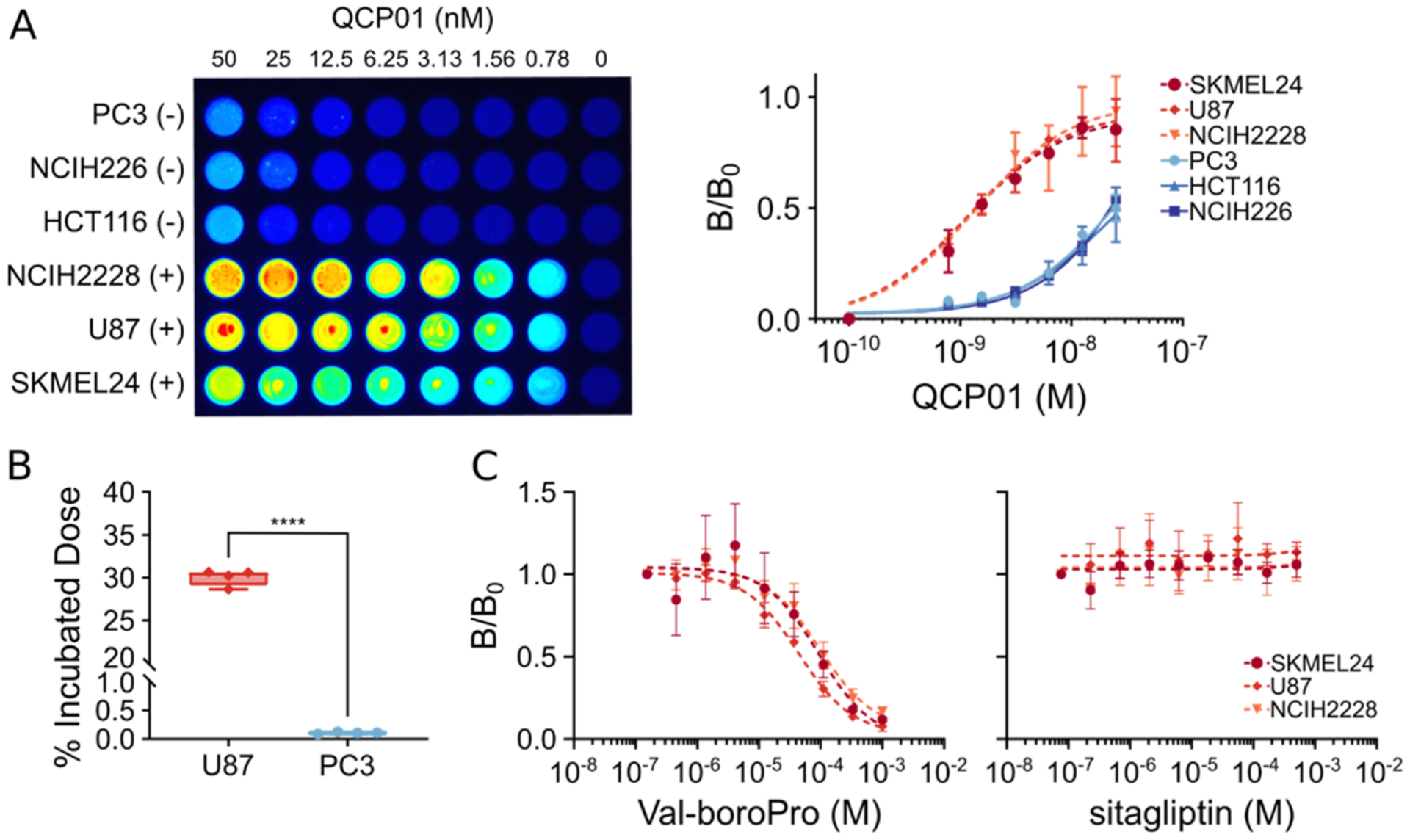
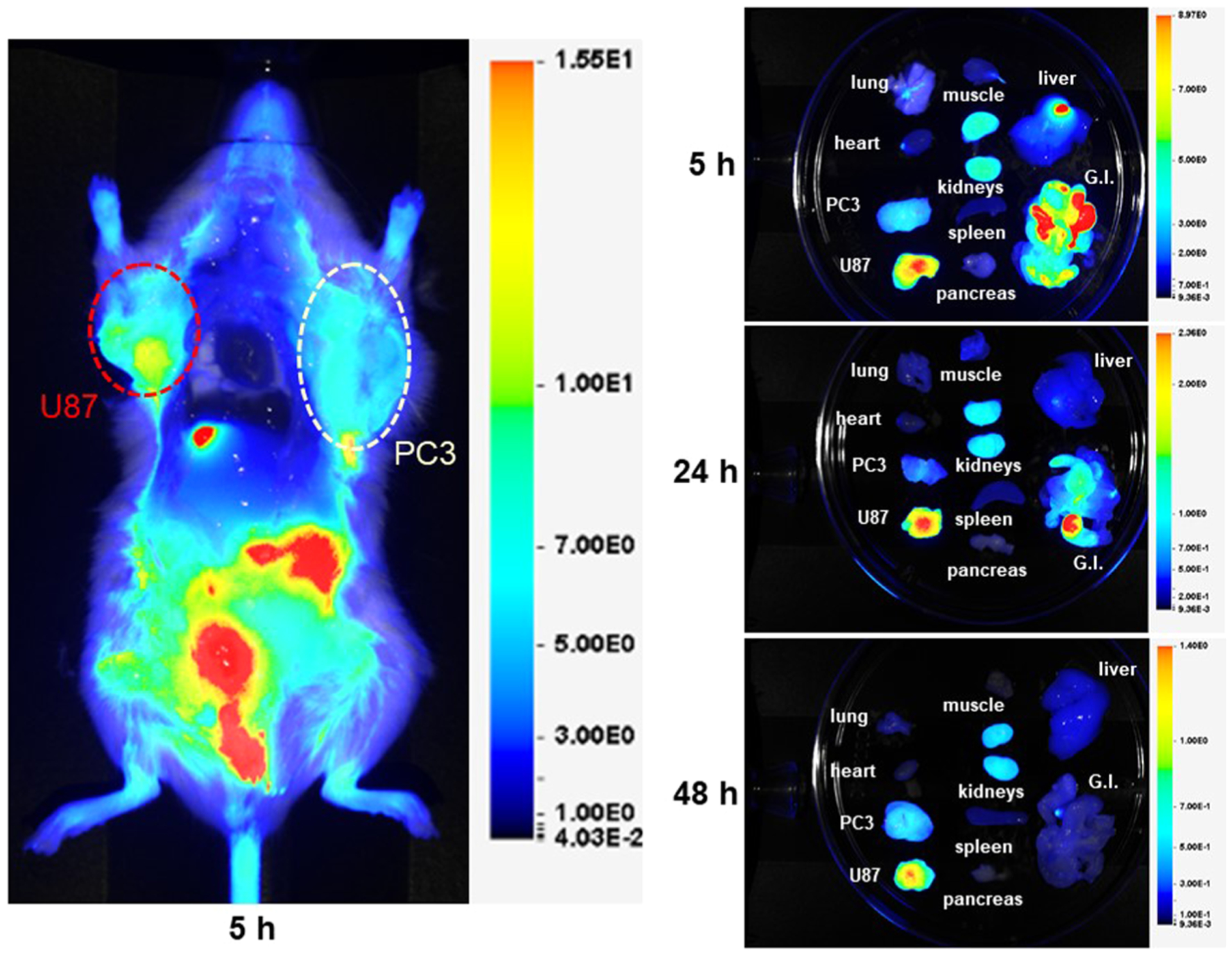
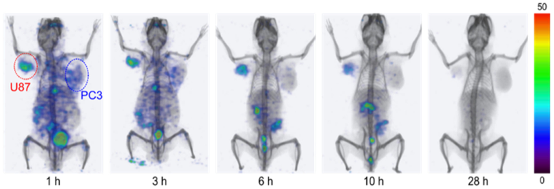
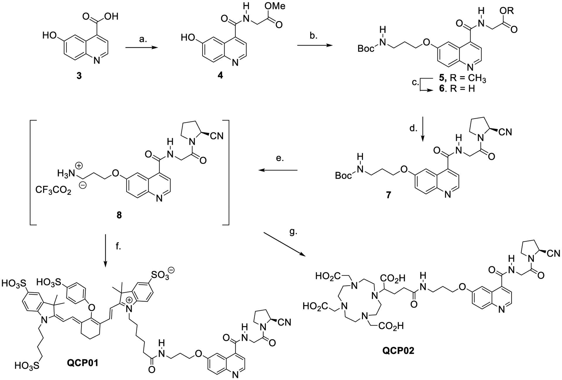
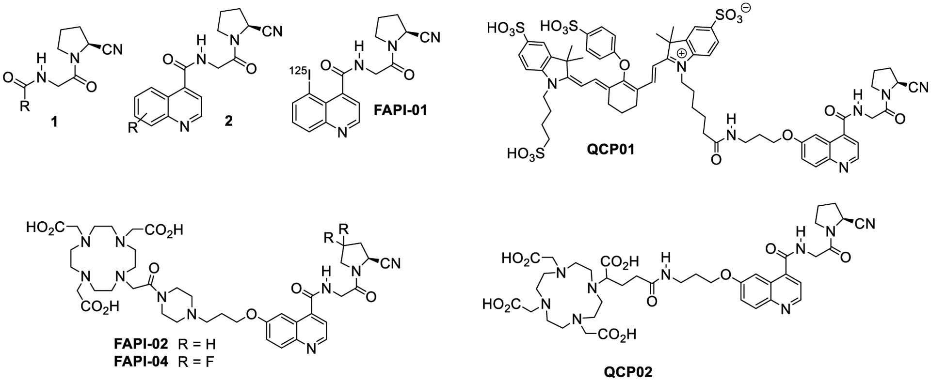
Fibroblast activation protein (FAP) has become a favored target for imaging and therapy of malignancy. We have synthesized and characterized two new (4-quinolinoyl)-glycyl-2-cyanopyrrolidine-based small molecules for imaging of FAP, QCP01 and [111In]QCP02, using optical and single-photon computed tomography/CT, respectively. Binding of imaging agents to FAP was assessed in six human cancer cell lines of different cancer types: glioblastoma (U87), melanoma (SKMEL24), prostate (PC3), NSCLC (NCIH2228), colorectal carcinoma (HCT116), and lung squamous cell carcinoma (NCIH226). Mouse xenograft models were developed with FAP-positive U87 and FAP-negative PC3 cells to test pharmacokinetics and binding specificity in vivo. QCP01 and [111In]QCP02 demonstrated nanomolar inhibition of FAP at Ki values of 1.26 and 16.20 nM, respectively. Both were selective for FAP over DPP-IV, a related serine protease. Both enabled imaging of FAP-expressing tumors specifically in vivo. [111In]QCP02 showed high uptake at 18.2 percent injected dose per gram in the U87 tumor at 30 min post-administration.
Figures
Similar articles
-
Rational modifications on N-(4-quinolinoyl)-Gly-2-cyanopyrrolidine to develop fibroblast activation protein-targeted radioligands with improved affinity and tumor uptake.Eur J Med Chem. 2025 Jan 5;281:117011. doi: 10.1016/j.ejmech.2024.117011. Epub 2024 Oct 29. Eur J Med Chem. 2025. PMID: 39488967
-
Extended structure-activity relationship and pharmacokinetic investigation of (4-quinolinoyl)glycyl-2-cyanopyrrolidine inhibitors of fibroblast activation protein (FAP).J Med Chem. 2014 Apr 10;57(7):3053-74. doi: 10.1021/jm500031w. Epub 2014 Mar 24. J Med Chem. 2014. PMID: 24617858
-
Targeting carcinoma-associated fibroblasts within the tumor stroma with a fibroblast activation protein-activated prodrug.J Natl Cancer Inst. 2012 Sep 5;104(17):1320-34. doi: 10.1093/jnci/djs336. Epub 2012 Aug 21. J Natl Cancer Inst. 2012. PMID: 22911669 Free PMC article.
-
Molecular recognition of fibroblast activation protein for diagnostic and therapeutic applications.Biochim Biophys Acta Proteins Proteom. 2020 Jul;1868(7):140409. doi: 10.1016/j.bbapap.2020.140409. Epub 2020 Apr 6. Biochim Biophys Acta Proteins Proteom. 2020. PMID: 32171757 Review.
-
Pro-tumorigenic roles of fibroblast activation protein in cancer: back to the basics.Oncogene. 2018 Aug;37(32):4343-4357. doi: 10.1038/s41388-018-0275-3. Epub 2018 May 3. Oncogene. 2018. PMID: 29720723 Free PMC article. Review.
Cited by
-
FAPI PET/CT Imaging-An Updated Review.Diagnostics (Basel). 2023 Jun 9;13(12):2018. doi: 10.3390/diagnostics13122018. Diagnostics (Basel). 2023. PMID: 37370912 Free PMC article. Review.
-
Experimental training in molecular pharmacology education based on drug-target interactions.Pharmacol Res Perspect. 2023 Aug;11(4):e01118. doi: 10.1002/prp2.1118. Pharmacol Res Perspect. 2023. PMID: 37548279 Free PMC article.
-
Expression of Fibroblast Activation Protein Is Enriched in Neuroendocrine Prostate Cancer and Predicts Worse Survival.Genes (Basel). 2022 Jan 13;13(1):135. doi: 10.3390/genes13010135. Genes (Basel). 2022. PMID: 35052475 Free PMC article.
-
Translating Molecules into Imaging-The Development of New PET Tracers for Patients with Melanoma.Diagnostics (Basel). 2022 Apr 29;12(5):1116. doi: 10.3390/diagnostics12051116. Diagnostics (Basel). 2022. PMID: 35626272 Free PMC article. Review.
-
Tumor microenvironment and fibroblast activation protein inhibitor (FAPI) PET: developments toward brain imaging.Front Nucl Med. 2023 Jul 18;3:1183471. doi: 10.3389/fnume.2023.1183471. eCollection 2023. Front Nucl Med. 2023. PMID: 39355017 Free PMC article. Review.
References
-
- Jacob M; Chang L; Pure E Fibroblast activation protein in remodeling tissues. Curr. Mol. Med 2012, 12, 1220–1243. - PubMed
-
- Miao Q; Yeo DC; Wiraja C; Zhang J; Ning X; Xu C; Pu K Near-Infrared Fluorescent Molecular Probe for Sensitive Imaging of Keloid. Angew. Chem., Int. Ed 2018, 57, 1256–1260. - PubMed
-
- van der Geest T; Roeleveld DM; Walgreen B; Helsen MM; Nayak TK; Klein C; Hegen M; Storm G; Metselaar JM; van den Berg WB; van der Kraan PM; Laverman P; Boerman OC; Koenders MI Imaging fibroblast activation protein to monitor therapeutic effects of neutralizing interleukin-22 in collagen-induced arthritis. Rheumatology 2018, 57, 737–747. - PubMed
-
- Yu DMT; Yao T-W; Chowdhury S; Nadvi NA; Osborne B; Church WB; McCaughan GW; Gorrell MD The dipeptidyl peptidase IV family in cancer and cell biology. FEBS J 2010, 277, 1126–1144. - PubMed
Publication types
MeSH terms
Substances
Grants and funding
LinkOut - more resources
Full Text Sources
Other Literature Sources
Medical
Miscellaneous
