. 2021 Jul 8:44:221-252.
doi: 10.1146/annurev-neuro-100520-012117. Epub 2021 Mar 17.

Neocortical Layer 1: An Elegant Solution to Top-Down and Bottom-Up Integration

Affiliations

Neocortical Layer 1: An Elegant Solution to Top-Down and Bottom-Up Integration

Benjamin Schuman et al. Annu Rev Neurosci. .

Abstract

Many of our daily activities, such as riding a bike to work or reading a book in a noisy cafe, and highly skilled activities, such as a professional playing a tennis match or a violin concerto, depend upon the ability of the brain to quickly make moment-to-moment adjustments to our behavior in response to the results of our actions. Particularly, they depend upon the ability of the neocortex to integrate the information provided by the sensory organs (bottom-up information) with internally generated signals such as expectations or attentional signals (top-down information). This integration occurs in pyramidal cells (PCs) and their long apical dendrite, which branches extensively into a dendritic tuft in layer 1 (L1). The outermost layer of the neocortex, L1 is highly conserved across cortical areas and species. Importantly, L1 is the predominant input layer for top-down information, relayed by a rich, dense mesh of long-range projections that provide signals to the tuft branches of the PCs. Here, we discuss recent progress in our understanding of the composition of L1 and review evidence that L1 processing contributes to functions such as sensory perception, cross-modal integration, controlling states of consciousness, attention, and learning.

Keywords: GABAergic interneurons; layer 1; neocortex; predictive coding; pyramidal cell dendrites; top-down processing.


Figures

Figure 1
Figure 1
The composition of neocortical L1. L1 is the only layer lacking excitatory cell somata. It contains several structures that mediate the integration of top-down and bottom-up information in PCs, the output neurons of the cortex located in deeper layers, via synapses on their distal dendritic tufts in L1. Top-down information is conveyed to L1 by dense axonal projections, including feedback projections from cortical areas higher in the processing hierarchy, axons from higher-order thalamocortical neurons mediating cortico-thalamo-cortical loops, and axons from neurons in neuromodulatory centers. L1 also contains a rich population of GABAergic INs, including four types of INs with somata in L1: NGFCs, α7, CanCs, and VIPs. L1 also contains a significant proportion of the dendrites of at least three populations of INs with somata in L2/3, including VIP/Bips, VIP/CCK, and ChCs. Lastly, L1 contains the ascending axon of SST-expressing Martinotti cells located in L2/3 and infragranular layers. L2/3 and L5—6 SST INs receive powerful local excitatory input, but so far they do not seem to receive long-range inputs. Abbreviations: α7, α7 nicotinic receptor–expressing interneuron; CanC, canopy cell; ChC, chandelier cell; IN, interneuron; L, layer; NGFC, neurogliaform cell; PC, pyramidal cell; SST, somatostatin; VIP, vasoactive intestinal peptide–expressing interneuron; VIP/Bip, vasoactive intestinal peptide–expressing bipolar interneuron; VIP/CCK, cholecystokinin- and vasoactive intestinal peptide–expressing interneuron.
Figure 2
Figure 2
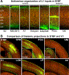
Laminar and sublaminar organization of L1 afferents in primary sensory cortex. (a) Corticocortical and thalamocortical inputs to the S1BF show specific laminar distributions and intriguing sublaminar biases within L1. Long-range cortical projections to L1 in S1BF include those from S2, M2 (also known as vM1), A1, and Ent/peri (see Section 3.5). In addition to these long-range corticocortical inputs, there is a distinct local projection to L1 from the subplate (L6b; CTGF+), with ascending collateral projections to L1 in M1/M2. Thalamocortical inputs include those from the higher-order POm and VM. These afferents exhibit specific laminar distributions and intriguing sublaminar biases within L1, with S2, vM1, and the subplate targeting superficial L1, while A1 and Ent/peri projections target mid/lower L1. The thalamocortical inputs from the POm and VM are also biased toward superficial L1. (b) Comparison of thalamocortical inputs to S1BF and V1 reveals specific trends in areal, laminar, and sublaminar distributions. Inputs from the first-order VPM and LGN relaying sensory information to S1 and V1, respectively, target L4 and L5b/6. In contrast, inputs from the higher-order POm to S1 and LP to V1 target L1 and L5a. Inputs from the higher-order VM and AV to S1 and V1, respectively, also target superficial L1 but lack the L5a projection. Moreover, their L1 projection is even more restricted in superficial L1 than the input from POm and LP. Projections from higher-order thalamic nuclei are diverse, with some exhibiting diffuse targeting of wide areas of the cortex like the VM, while others are more specific, such as the POm with projections largely restricted to somatosensory/motor areas (see insets). The laminar, sublaminar, and areal specificity of corticocortical and thalamocortical projections illustrated here likely contribute to the specific organization and processing of distinct inputs along pyramidal neuron apical dendrites. The widespread innervation of cortical areas by the VM might explain its role in global arousal (see Section 3.3). Abbreviations: A1, primary auditory cortex; AV, anteroventral thalamic nucleus; Ent/peri, entorhinal/perirhinal cortex; L, layer; LGN, lateral geniculate nucleus; LP, lateral posterior thalamic nucleus; M1, primary motor cortex; M2, secondary motor cortex; POm, posteromedial thalamic nucleus; S1, primary somatosensory cortex; S1BF, somatosensory barrel field cortex; S2, secondary somatosensory cortex; V1, primary visual cortex; VM, ventromedial thalamic nucleus; vM1, vibrissal motor cortex; VPM, ventroposteromedial thalamic nucleus. Images of fluorescently labeled projections are from the Allen Institute (http://connectivity.brain-map.org)
Figure 3
Figure 3
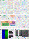
Properties of the four resident GABAergic INs of L1. The four IN subtypes (NGFCs, canopy cells, α7s, and VIPs) have unique morphological and electrophysiological properties. (a) Voltage responses of L1 INs (middle traces in color) in response to hyperpolarizing, just-subthreshold, just-threshold, and suprathreshold current injections (black; bottom traces). Insets show the near-threshold depolarizing hump in α7 cells that is absent in canopy cells. Note the near-threshold delayed spiking in NGFCs, high input resistance in VIP cells, and higher spike frequency adaption for α7 and VIP cells. Main scale bars are 20mV or 400pA and 200ms; inset scale bars are 15mV and 40ms. (b) Morphological reconstructions of the four resident IN populations. NGFCs and canopy cells have their axon largely confined to L1 spanning several columns, while α7 and VIP INs have descending translaminar axonal collaterals. Inset shows multipolar dendrites of α7 cells and bipolar dendrites of VIP cells. (c) Molecular markers segregate the four resident L1 INs. NGFCs and canopy cells both express NDNF, but NGFCs also express NPY. Non-NDNF IN populations include the α7 and the VIP INs. (d) The density of GABAergic INs in L1 is similar to that of other cortical layers. (Left) The cortical column in primary somatosensory cortex is stained for Nissl in a GAD67-GFP mouse. (Middle) GFP in the same cortical column is shown. (Right) The density of GAD67-expressing neurons across cortical layers (N = 2,656 counted cells) is shown. (e) NGFCs and canopy cells have similar morphologies (panel b) but differ in electrophysiological properties (panel a), connectivity (see text and Schuman et al. 2019), and cholinergic responses. The effects of a 30-ms puff of 20 μM muscarine (blue arrow) as the cells were depolarized to produce an approximately 5–10-Hz spike train are shown. A representative trace is shown in black, and all traces from that session are shown in gray. Abbreviations: α7, α7 nicotinic receptor–expressing interneuron; GFP, green fluorescent protein; IN, interneuron; L, layer; nAChR, nicotinic acetylcholine receptor; NDNF, neuron-derived neurotrophic factor; NGFC, neurogliaform cell; NPY, neuropeptide Y; VIP, vasoactive intestinal peptide–expressing interneuron. Panels a, b, and c modified from Schuman et al. (2019).

Similar articles

Cited by

References

    1. Abs E, Poorthuis RB, Apelblat D, Muhammad K, Pardi MB, et al. 2018. Learning-related plasticity in dendrite-targeting layer 1 interneurons. Neuron 100:684–99.e6 - PMC - PubMed
    1. Adesnik H, Bruns W, Taniguchi H, Huang ZJ, Scanziani M. 2012. A neural circuit for spatial summation in visual cortex. Nature 490:226–31 - PMC - PubMed
    1. Allaway KC, Muñoz W, Tremblay R, Sherer M, Herron J, et al. 2020. Cellular birthdate predicts laminar and regional cholinergic projection topography in the forebrain. eLife 9:e63249 - PMC - PubMed
    1. Anastasiades PG, Collins DP, Carter AG. 2020. Mediodorsal and ventromedial thalamus engage distinct L1 circuits in the prefrontal cortex. Neuron 109:314–30.e4 - PMC - PubMed
    1. Arezzo JC, Vaughan HG Jr., Legatt AD. 1981. Topography and intracranial sources of somatosensory evoked potentials in the monkey. II. Cortical components. Electroencephalogr. Clin. Neurophysiol 51:1–18 - PubMed

Publication types

LinkOut - more resources