. 2021 Mar 16;34(11):108869.
doi: 10.1016/j.celrep.2021.108869.

The mitochondrial carrier SFXN1 is critical for complex III integrity and cellular metabolism

Affiliations

The mitochondrial carrier SFXN1 is critical for complex III integrity and cellular metabolism

Michelle Grace Acoba et al. Cell Rep. .

Abstract

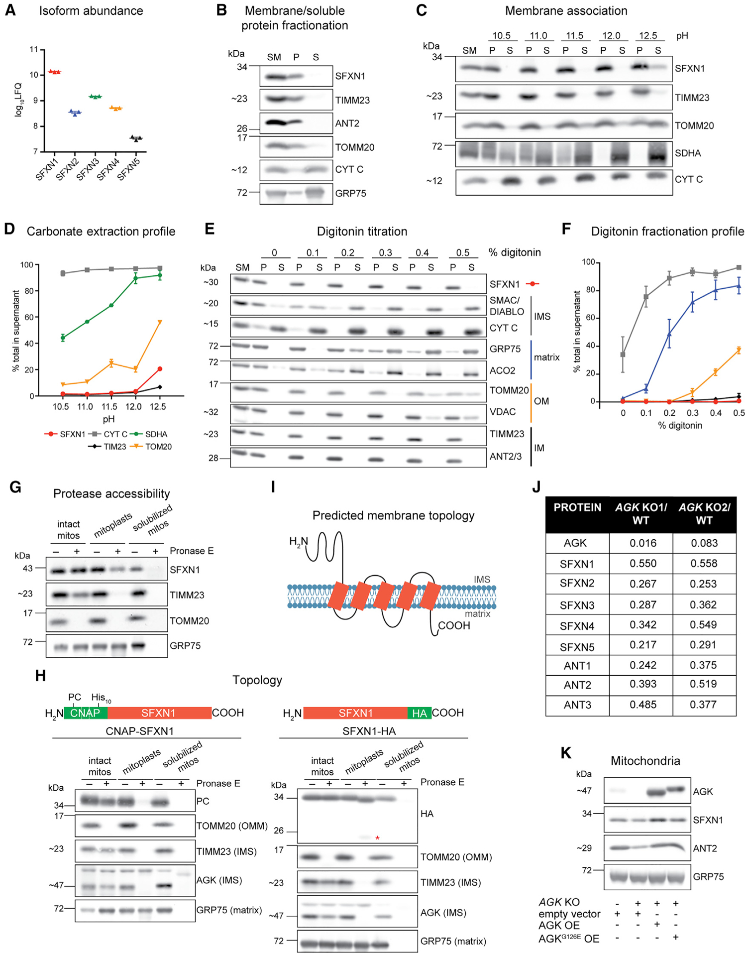
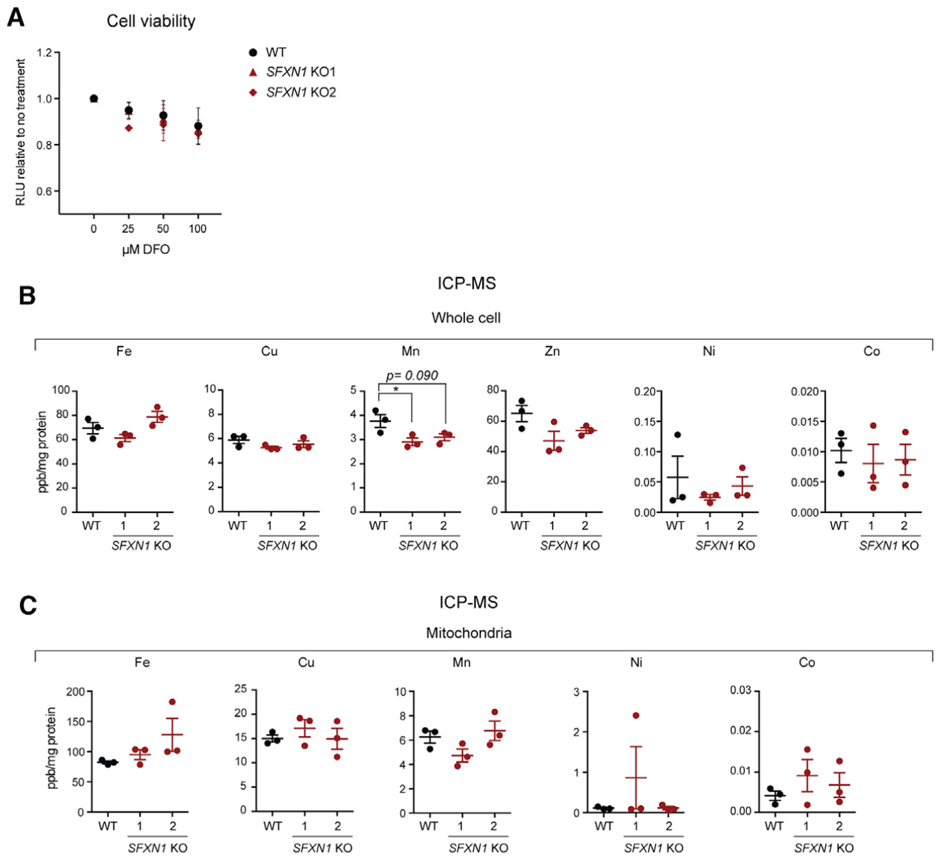
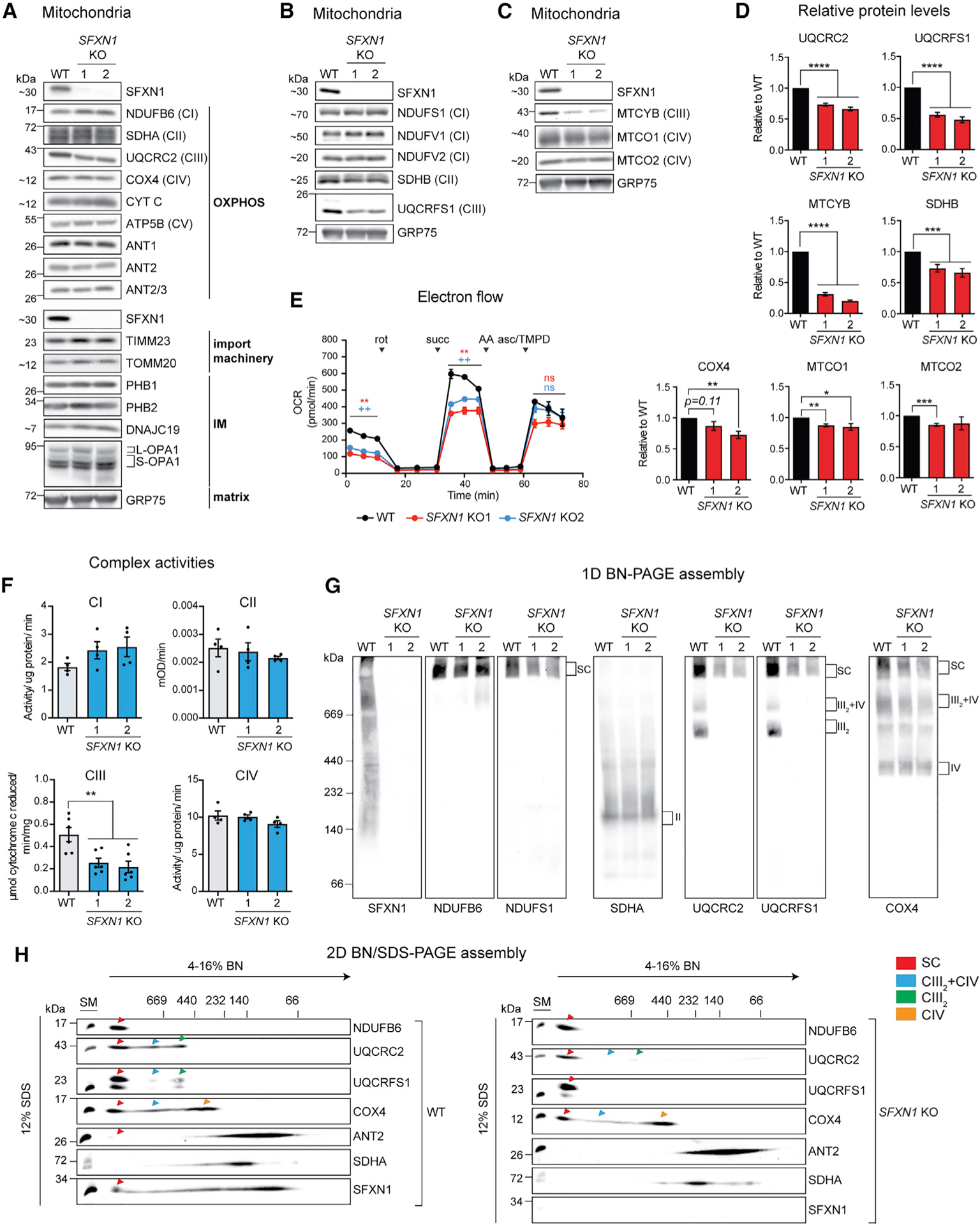
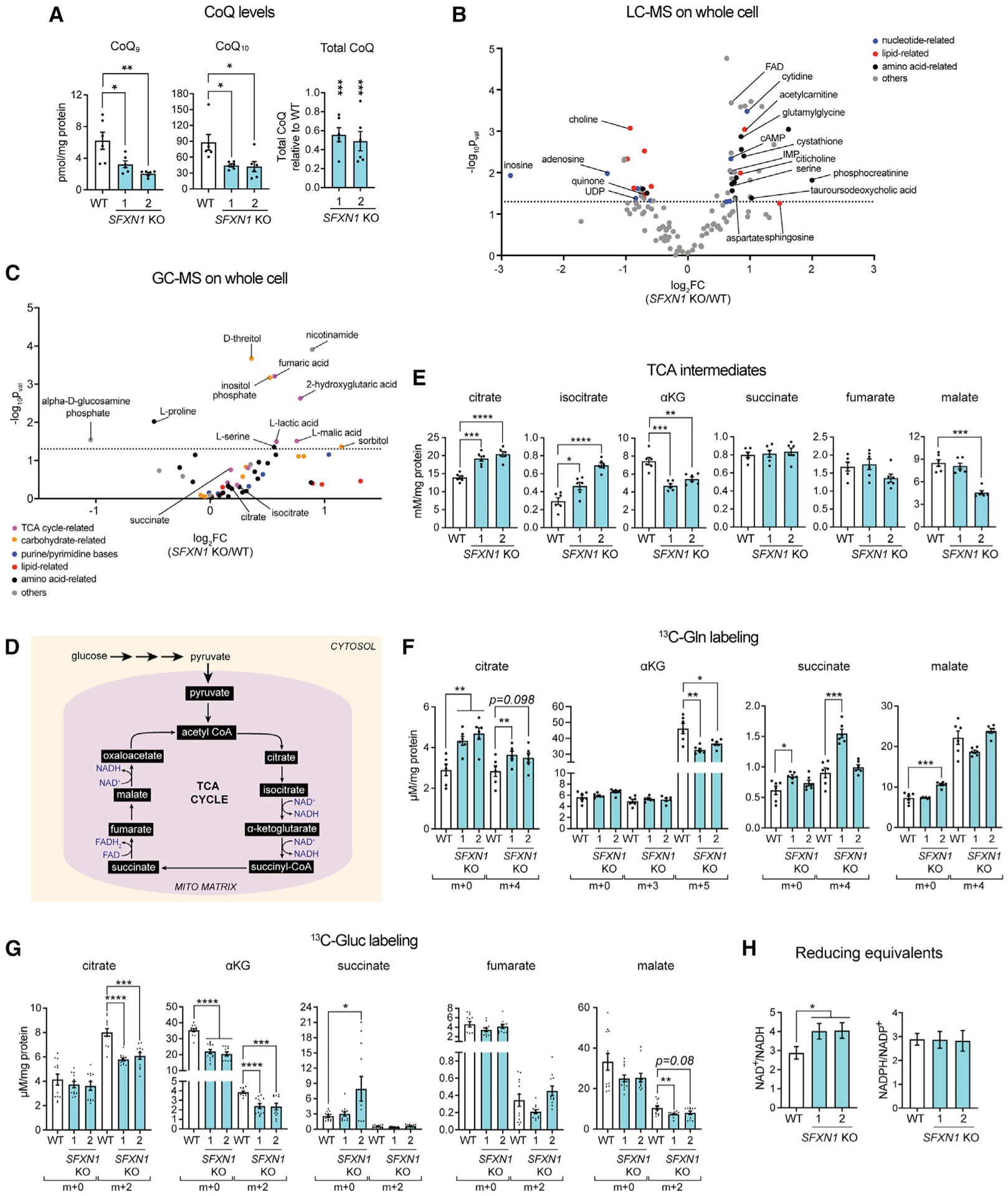
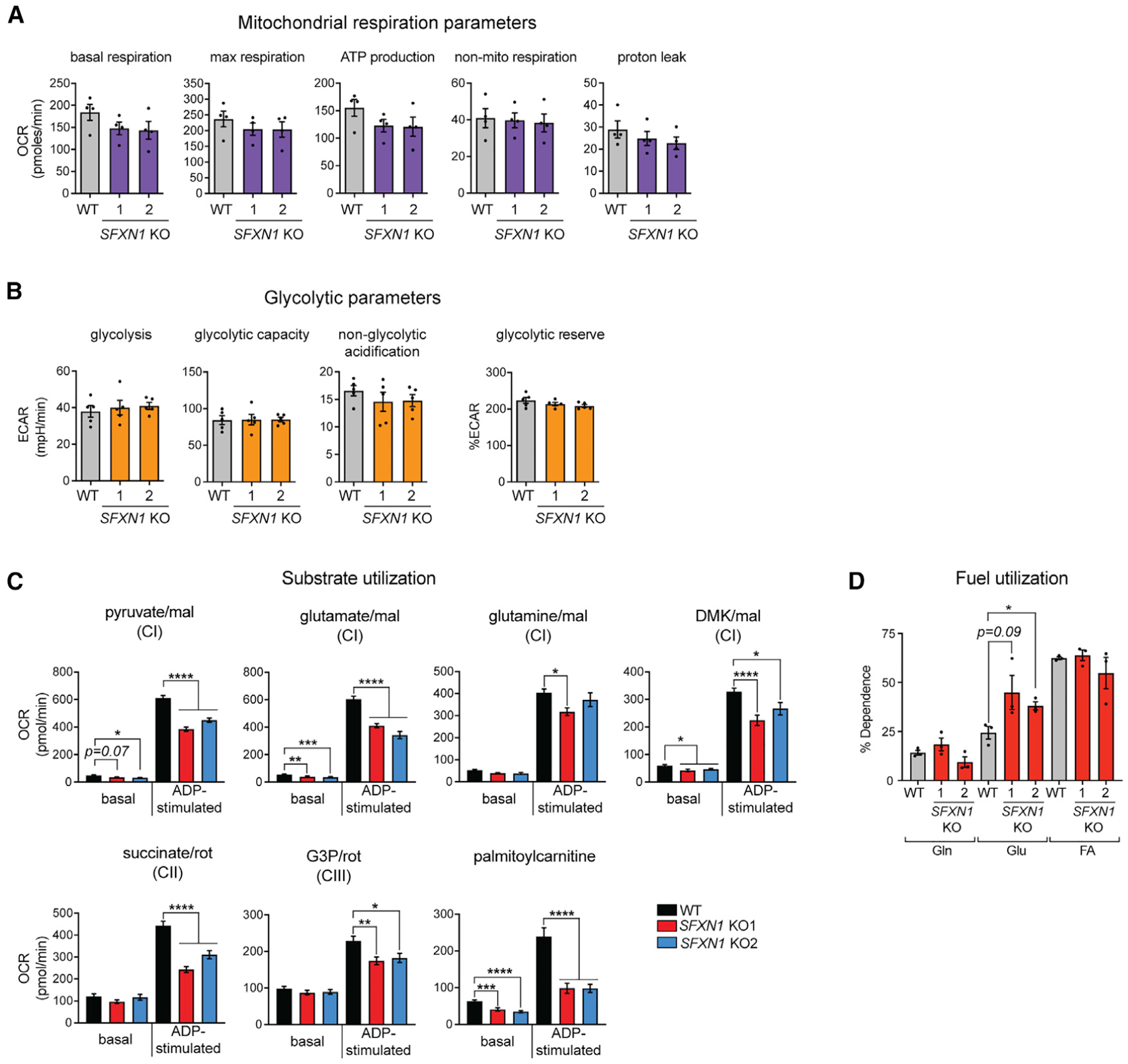
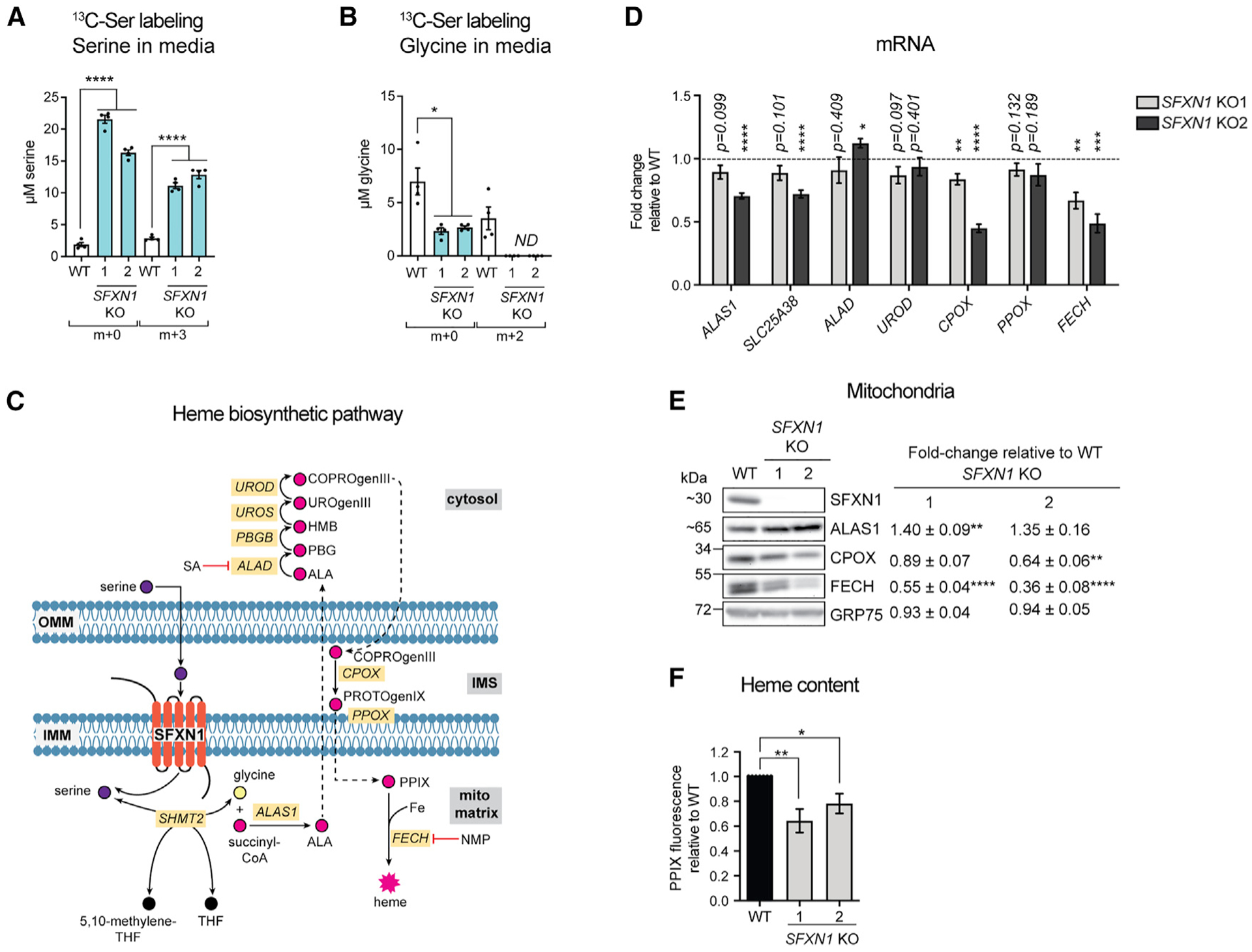
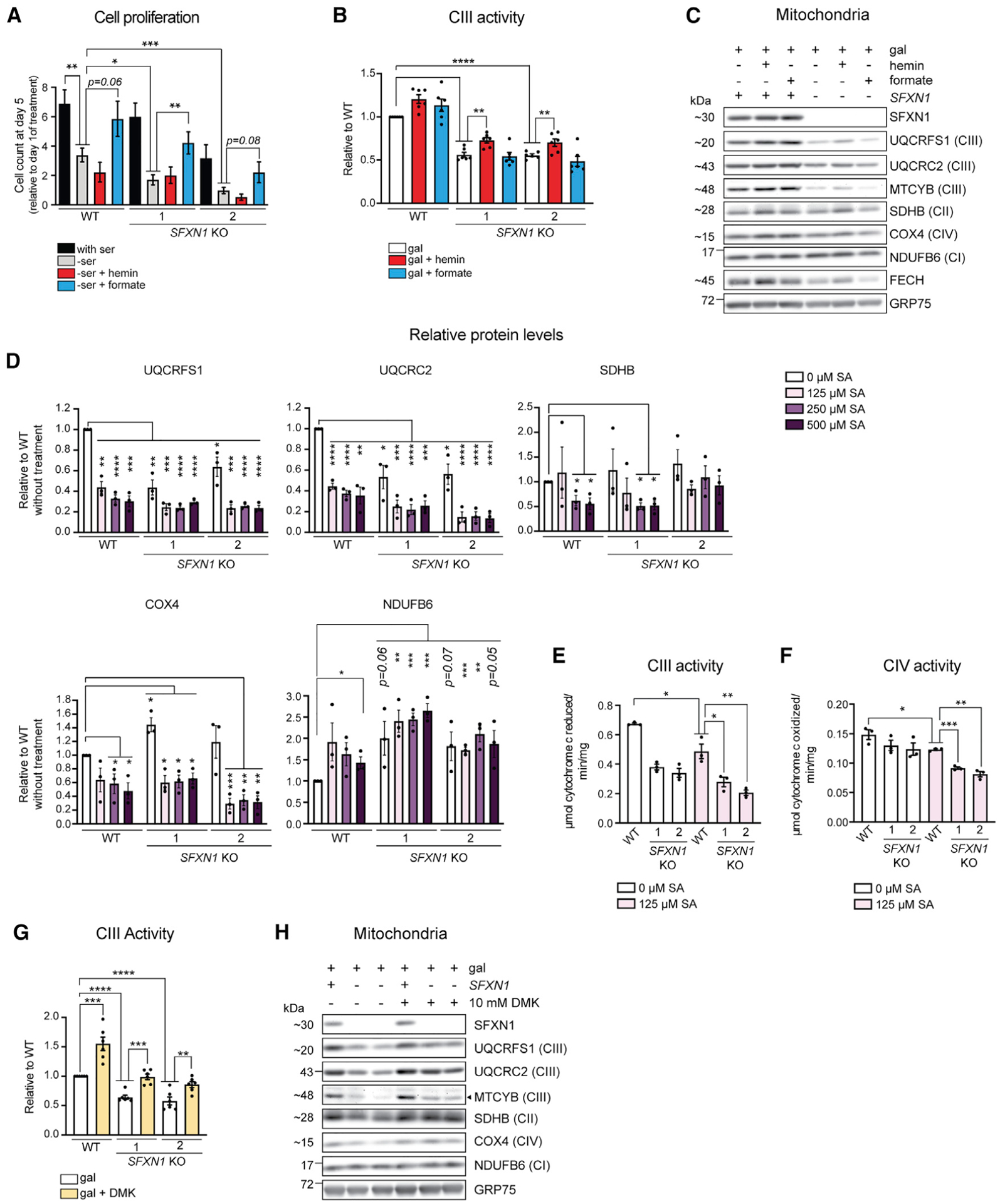
Mitochondrial carriers (MCs) mediate the passage of small molecules across the inner mitochondrial membrane (IMM), enabling regulated crosstalk between compartmentalized reactions. Despite MCs representing the largest family of solute carriers in mammals, most have not been subjected to a comprehensive investigation, limiting our understanding of their metabolic contributions. Here, we functionally characterize SFXN1, a member of the non-canonical, sideroflexin family. We find that SFXN1, an integral IMM protein with an uneven number of transmembrane domains, is a TIM22 complex substrate. SFXN1 deficiency leads to mitochondrial respiratory chain impairments, most detrimental to complex III (CIII) biogenesis, activity, and assembly, compromising coenzyme Q levels. The CIII dysfunction is independent of one-carbon metabolism, the known primary role for SFXN1 as a mitochondrial serine transporter. Instead, SFXN1 supports CIII function by participating in heme and α-ketoglutarate metabolism. Our findings highlight the multiple ways that SFXN1-based amino acid transport impacts mitochondrial and cellular metabolic efficiency.

Keywords: Complex III; OXPHOS; SFXN1; TIM22 complex; amino acid; heme; mitochondria; mitochondrial carrier; serine; sideroflexin.


Conflict of interest statement

Declaration of interests The authors declare no competing interests.

Figures

Figure 1.
Figure 1.. SFXN1, an integral inner mitochondrial membrane protein, is a TIM22 complex substrate
(A) Relative protein abundance of SFXN isoforms in HEK293 mitochondria as, determined by mass spectrometry and label-free quantification (LFQ) (mean ± SEM, n = 3). (B) Sonication and centrifugation of mitochondria to separate membrane-bound from soluble proteins. SM, starting material; P, pellet; S, supernatant. (C) Carbonate extraction of mitochondrial membrane proteins to distinguish between peripheral (appear in S) and integral (remain mostly in P) proteins. (D) Band intensities of P and S fractions in (C) were quantified and plotted as % of protein released in the supernatant (mean ± SEM, n = 3). (E) Digitonin titration for fractionation of mitochondrial subcompartments. Equal volumes of P and S fractions were analyzed. (F) Band intensities of P and S fractions (E) were quantified. Average band intensity of representative mitochondrial proteins from each subcompartment was plotted as % of protein released in the supernatant (mean ± SEM, n = 3). (G) Submitochondrial localization of endogenous SFXN1. HEK293 mitochondria were osmotically ruptured to yield mitoplasts or solubilized with sodium deoxycholate. Samples were treated with Pronase E where indicated. (H) Submitochondrial localization of tagged SFXN1. HEK293 mitochondria lacking endogenous SFXN1 and expressing CNAP-SFXN1 or SFXN1-HA were processed as in (G). *, matrix-protected fragment. (I) Predicted membrane topology of SFXN1 based on (H). (J) Proteomic analysis of AGK KOs versus WT. Shown are relative protein amounts of SFXN and ANT isoforms in the presence or absence of AGK. (K) Mitochondria from AGK KOs rescued with AGK, AGKG126E, or empty vector were resolved by SDS-PAGE and immunoblotted for the indicated proteins.
Figure 2.
Figure 2.. Deletion of SFXN1 alone does not disrupt iron metabolism
(A) Cell viability of HEK293 WT and SFXN1 KOs upon treatment with the indicated concentration of the iron chelator deferoxamine mesylate (DFO) for 24 h (mean ± SEM, n = 3). (B and C) Relative metal concentrations in WT and SFXN1 KO cells (B) and mitochondria (C), as determined by ICP-MS (mean ± SEM, n = 3). *p < 0.05; unpaired Student’s t test.
Figure 3.
Figure 3.. Absence of SFXN1 leads to complex-III-related defects
(A) Steady-state abundance of select mitochondrial proteins, including OXPHOS components, subunits of import machineries, and other proteins in the IMM and matrix. (B) Immunoblotting for Fe-S containing subunits of respiratory complexes in mitochondrial extracts. GRP75 served as loading control. (C) Immunoblotting for mtDNA-encoded subunits in mitochondrial extracts. GRP75 served as loading control. (D) Densitometric analysis of bands for select proteins in (A), (B), and (C). Protein levels in WT were set to 1.0 (mean ± SEM, n ≥ 4). (E) Electron flow through the respiratory chain in intact mitochondria. Base buffer contains 10 mM pyruvate, 2 mM malate, and 4 μM carbonyl cyanide m-chlorophenyl hydrazone (CCCP). Injections of complex inhibitors/substrates were performed as indicated. rot, rotenone; succ, succinate; AA, antimycin A; asc/TMPD, ascorbate/N,N,N′,N′-tetramethyl-p-phenylenediamine. (F) Spectrophotometric assays using detergent-solubilized mitochondria to monitor individual complex activities. CI, oxidation of NADH to NAD+; CII, ubiquinol production; CIII, cytochrome c reduction; CIV, cytochrome c oxidation (mean ± SEM, n ≥ 4). (G) 1D BN assembly. Mitochondria solubilized in 1% (w/v) digitonin were resolved on a 4%–16% BN gel and immunoblotted for the indicated subunits. (H) Mitochondria solubilized in 1% (w/v) digitonin were resolved by 2D BN/SDS-PAGE and immunoblotted for the indicated subunits. *p < 0.05, **p < 0.01, ***p < 0.001, ****p < 0.0001; unpaired Student’s t test.
Figure 4.
Figure 4.. SFXN1 loss results in metabolic perturbations
(A) CoQ measurements in cellular lipid isolates by reversed phase high-performance liquid chromatography-MS (RP-HPLC/MS) (mean ± SEM, n = 6). (B and C) Metabolite abundance in whole cells obtained by untargeted LC-MS (B) or GC-MS (C) analysis (n = 6 per group). (D) TCA cycle. (E) Steady-state intracellular concentrations of TCA cycle metabolites obtained by LC-MS/MS (mean ± SEM, n = 6). αKG, α-ketoglutarate. (F and G) Levels of m+x-labeled metabolites as determined by LC-MS/MS upon [U-13C]-glutamine labeling (mean ± SEM, n = 6) (F) and [U-13C]-glucose labeling (mean ± SEM, n = 12) (G). (H) Quantification of NAD+/NADH (mean ± SEM, n ≥ 8) and NADPH/NADP+ ratios (mean ± SEM, n = 6). *p < 0.05, **p < 0.01, ***p < 0.001, ****p < 0.0001; unpaired Student’s t test.
Figure 5.
Figure 5.. Cells lacking SFXN1 display metabolic flexibility
(A) Oxygen consumption rate (OCR) in intact cells. Presented are values normalized by DNA content (mean ± SEM, n = 4). (B) Extracellular acidification rate (ECAR) in intact cells. Presented are values normalized by DNA content (mean ± SEM, n = 5). (C) OCR in isolated mitochondria using the specified substrates (mean ± SEM, n ≥ 9 wells from at least 3 independent experiments). mal, malate; DMK, dimethyl αKG; G3P, glycerol-3-phosphate. (D) Percent dependency on glutamine (Gln), glucose (Gluc), and fatty acid (FA) use (mean ± SEM, n = 3). *p < 0.05, **p < 0.01, ***p < 0.001, ****p < 0.0001; unpaired Student’s t test.
Figure 6.
Figure 6.. Heme biosynthesis is compromised in SFXN1 Kos
(A and B) Quantification of m+x serine (A) and m+x glycine (B) in media by LC-MS/MS. Cells were grown in serine- and glycine-free media with [U]-13C-serine for 24 h (mean ± SEM, n = 4). (C) Heme biosynthetic pathway. SA, succinylacetone; NMP, N-methyl protoporphyrin; PPIX, protoporphyrin IX. (D) Gene expression analysis of heme biosynthetic enzymes by qPCR (mean fold-change [FC] ± SEM, n = 4). (E) Immunoblotting for enzymes involved in heme biosynthesis. Values shown are fold-change protein steady-state abundance in SFXN1 KOs over HEK293 WT (mean ± SEM, n = 5). (F) Relative total heme content obtained by analyzing total heme fluorescence (mean ± SEM, n = 7). *p < 0.05, **p < 0.01, ***p < 0.001, ****p < 0.0001; unpaired Student’s t test.
Figure 7.
Figure 7.. Reinforcement of heme and αKG metabolism, but not the one-carbon pathway, partially restores complex III function in the absence of SFXN1
(A) Cell proliferation in full media (with serine), without serine (-ser), and upon supplementation of serine-free media with 15 μM hemin (−ser + hemin) or 1 mM formate (−ser + formate) (mean ± SEM, n ≥ 4). (B) Complex III activity in DDM-solubilized mitochondria. At 48 h before mitochondrial isolation, cells were switched to media containing galactose only, galactose with 15 μM hemin, or galactose with 1 mM formate (mean ± SEM, n = 6). (C) Immunoblotting for select respiratory complex subunits using mitochondrial isolates detailed in (B). (D) Steady-state protein abundance of select respiratory complex subunits in cell lysates. Cells were grown in glucose-based media and treated with the indicated concentration of SA, an inhibitor of heme biosynthesis, for 2 days. (E and F) Complex III (E) or IV (F) activity in DDM-solubilized mitochondria. At 48 h before mitochondrial isolation, cells were switched to media containing galactose only or galactose with 125 μM SA (mean ± SEM, n = 3). (G) Complex III activity in DDM-solubilized mitochondria. At 48 h before mitochondrial isolation, cells were switched to media containing galactose only or galactose with 10 mM dimethyl αKG (DMK) (mean ± SEM, n = 6). (H) Immunoblotting for select respiratory complex subunits using mitochondrial isolates detailed in (G). *p < 0.05, **p < 0.01, ***p < 0.001, ****p < 0.0001; unpaired Student’s t test.

References

    1. Abrahams JP, Leslie AG, Lutter R, and Walker JE (1994). Structure at 2.8 A resolution of F1-ATPase from bovine heart mitochondria. Nature 370, 621–628. - PubMed
    1. Adam A, Endres M, Sirrenberg C, Lottspeich F, Neupert W, and Brunner M (1999). Tim9, a new component of the TIM22.54 translocase in mitochondria. EMBO J 18, 313–319. - PMC - PubMed
    1. Ajioka RS, Phillips JD, and Kushner JP (2006). Biosynthesis of heme in mammals. Biochim. Biophys. Acta 1763, 723–736. - PubMed
    1. Al Kadhi O, Melchini A, Mithen R, and Saha S (2017). Development of a LC-MS/MS Method for the Simultaneous Detection of Tricarboxylic Acid Cycle Intermediates in a Range of Biological Matrices. J. Anal. Methods Chem 2017, 5391832. - PMC - PubMed
    1. Amelio I, Cutruzzolá F, Antonov A, Agostini M, and Melino G (2014). Serine and glycine metabolism in cancer. Trends Biochem. Sci 39, 191–198. - PMC - PubMed

Publication types

MeSH terms