. 2021 Mar 17;26(1):11.
doi: 10.1186/s11658-021-00255-y.

Triazoloacridone C-1305 impairs XBP1 splicing by acting as a potential IRE1α endoribonuclease inhibitor

Affiliations

Triazoloacridone C-1305 impairs XBP1 splicing by acting as a potential IRE1α endoribonuclease inhibitor

Sylwia Bartoszewska et al. Cell Mol Biol Lett. .

Abstract

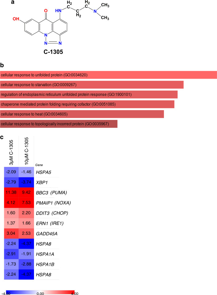
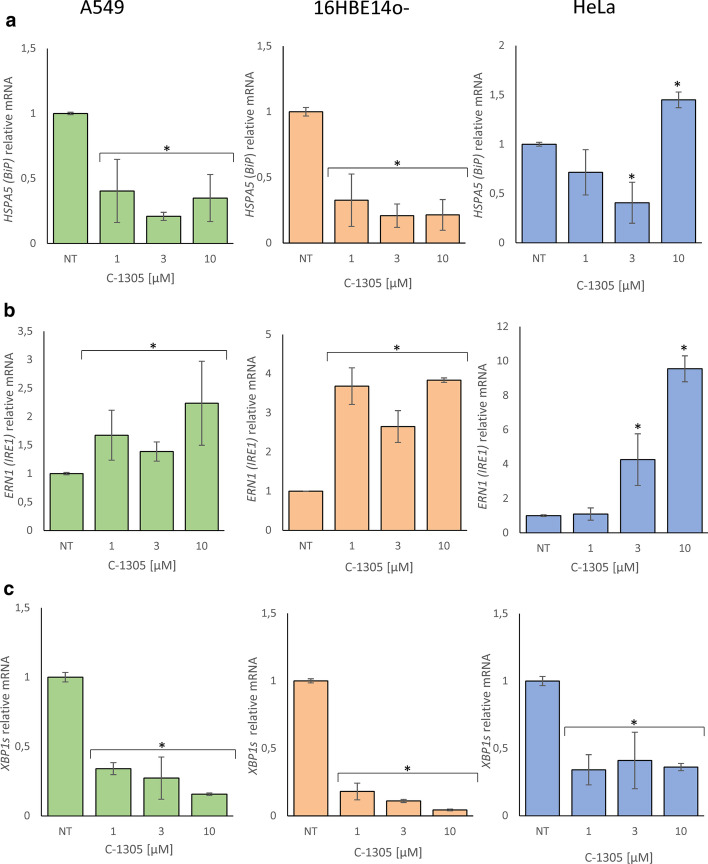
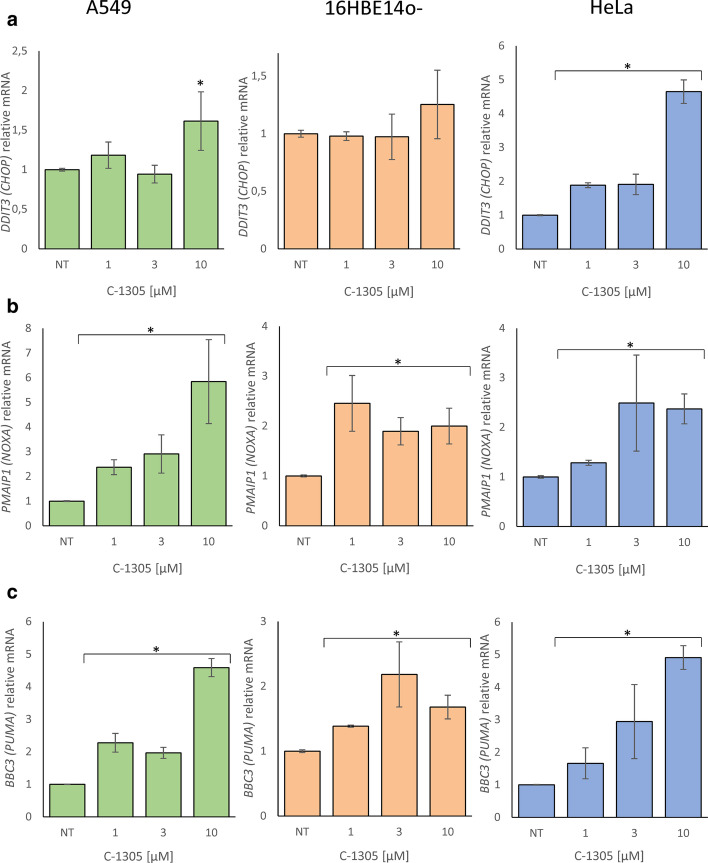
Inositol requiring enzyme 1 alpha (IRE1α) is one of three signaling sensors in the unfolding protein response (UPR) that alleviates endoplasmic reticulum (ER) stress in cells and functions to promote cell survival. During conditions of irrevocable stress, proapoptotic gene expression is induced to promote cell death. One of the three signaling stressors, IRE1α is an serine/threonine-protein kinase/endoribonuclease (RNase) that promotes nonconventional splicing of XBP1 mRNA that is translated to spliced XBP1 (XBP1s), an active prosurvival transcription factor. Interestingly, elevated IRE1α and XBP1s are both associated with poor cancer survival and drug resistance. In this study, we used next-generation sequencing analyses to demonstrate that triazoloacridone C-1305, a microtubule stabilizing agent that also has topoisomerase II inhibitory activity, dramatically decreases XBP1s mRNA levels and protein production during ER stress conditions, suggesting that C-1305 does this by decreasing IRE1α's endonuclease activity.

Keywords: ER stress; IRE1α; UPR; XBP1s.


Conflict of interest statement

The authors declare no conflict of interest. The funders had no role in the design of the study; in the collection, analyses, or interpretation of data; in the writing of the manuscript, or in the decision to publish the results.

Figures

Fig. 1
Fig. 1
The NGS and pathway analyses of early gene transcripts dysregulated by C-1305 treatment in A549 cells. a The 2D chemical structure of C-1305 (5-((3-(dimethylamino)propyl)amino)-8-hydroxy-6H-[1,2,3]triazolo[4,5,1-de]acridin-6-one; C18H19N5O2). b The unique early gene transcripts dysregulated after 8 h of treatment of A549 with 3 µM C-1305 were selected from the NGS experiments. Well-established gene transcripts with greater than 10 RPKMs per sample and with significance (p ≤ 0.05) greater for change in expression between C-1305-treated and control groups (no treatment and 24-h treatment) were used in pathway analyses. The Gene Ontology clusters are depicted for selected genes; clusters are listed followed by the p values and enrichment scores calculated by Enricher, which is used to determine the percentage of the chart. The longer bar the lower p-value, while the darker the color, the more enriched the cluster. Only clusters with p ≤ 0.001 were considered. c The heat map representing C-1305 exposure related expression changes in genes related to UPR and ER stress as observed in NGS experiments in A549 cells exposed to 3 µM and 10 µM C-1305 for 8 h. Heat maps were generated with the Morpheus software (Morpheus, https://software.broadinstitute.org/morpheus). The color scale and values depict fold change (c)
Fig. 2
Fig. 2
C-1305-induced changes in a BiP, b ERN1, c XBP1s mRNA levels in A549, 16HBE14o- and HeLa cells after 8 h of treatment. The results from 3 independent experiments (n = 9) are plotted normalized to GAPDH and TBP mRNA levels and expressed as a fold-change over the vehicle controls. Error bars represent standard deviations. Significant changes (p-value < 0.05) are marked with an asterisk
Fig. 3
Fig. 3
C-1305-induced changes in a CHOP, b NOXA, c PUMA mRNA levels in A549, 16HBE14o- and HeLa cells after 8 h of treatment. The results from 3 independent experiments (n = 9) are plotted normalized to GAPDH and TBP mRNA levels and expressed as a fold-change over the vehicle controls. Error bars represent standard deviations. Significant changes (p-value < 0.05) are marked with an asterisk
Fig. 4
Fig. 4
C-1305-prevents IRE1α-dependent XBP1 mRNA splicing during ER stress. a The 2D chemical structure of 4µ8C (7-Hydroxy-4-methyl-2-oxo-2H-chromene-8-carbaldehyde, C11H8O4). b HeLa cells were treated with ER stressors (Tm, 0.5 µg/ml) for 6 h in a presence of specified concentrations of C-1305 and 4µ8C. Following the treatments, the XBP1s mRNA levels were accessed with qPCR and expressed as a fold-change over the no stress controls. The results from 3 independent experiments (n = 9) are plotted normalized to GAPDH and TBP mRNA. Error bars represent standard deviations. Significant changes (p-value < 0.05) are marked with an asterisk. c The corresponding changes in XBP1s protein levels were evaluated by Western blot (d) normalized to total protein levels and related no stress control. The data from 3 independent experiments were analyzed. *p < 0.05 was considered significant. The raw data are provided in Additional file 1
Fig. 5
Fig. 5
The XBP1s mRNA-based determination of C-1305 impact on IRE1α endoribonuclease activity in ER stressed HeLa cells. HeLa cells were treated with ER stressors (Tm, 0.5 µg/ml and Tg, 25 nM), for 6 h in a presence of specified concentrations of C-1305 and 4µ8C. Following the treatments, the XBP1s mRNA levels were accessed with qPCR and expressed as a change versus ER stressed cells treated with vehicle. The results from 3 independent experiments (n = 9) are plotted normalized to GAPDH and TBP mRNA. Error bars represent standard deviations. Concentration response curves and IC50 values of a C-1305 and b 4µ8C in Tm treated cells were determined using Sigma Plot 1.1 software. Similar approach was used to determine Concentration response curves and IC50 values of c C-1305 and d 4µ8C in Tg treated cells. The IRE1α kinase assays were performed in the presence of serial dilution and were measured with ADP-Glo kinase assay. Concentration response curve and IC50 values of e C-1305, f 4µ8C and g staurosporine (used as a control of kinase activity) were determined using Origin Pro software. The results were expressed as remaining activity, normalized to the uninhibited control for 100% activity and to the fully inhibited one for 0% activity as described in “Materials and methods”. Data are plotted as means ± SD from triplicate measurements of two independent measurements (n = 6)
Fig. 6
Fig. 6
Prediction of C-1305, 4µ8C, and 3,6-DMAD binding mode in the and in the endoribonuclease (RNase) domain of IRE1α. The binding mode of a C-1305, b 4µ8C, and c 3,6-DMAD was modeled using the EDock server (https://zhanglab.ccmb.med.umich.edu/EDock/). The native and modeled structures of the IRE1 chain A are represented in green and yellow, respectively. ADP structure is represented by red, C-1305 by red sticks, 4µ8C by pink sticks, and 3,6-DMAD by sandy brown sticks. Predicting protein–ligand binding residues are represented by grey sticks together with oxygen, nitrogen, sulfur, and hydrogen atoms (colored red, blue, yellow, and white, respectively) and numbered according to IRE1α sequence (PDB ID code 4YZD). Magnesium ion (Mg) is colored by cornflower blue

Similar articles

Cited by

References

    1. Madden E, Logue SE, Healy SJ, Manie S, Samali A. The role of the unfolded protein response in cancer progression: from oncogenesis to chemoresistance. Biol Cell. 2019;111(1):1–17. doi: 10.1111/boc.201800050. - DOI - PubMed
    1. Schroder M, Kaufman RJ. ER stress and the unfolded protein response. Mutat Res. 2005;569(1–2):29–63. doi: 10.1016/j.mrfmmm.2004.06.056. - DOI - PubMed
    1. Sano R, Reed JC. ER stress-induced cell death mechanisms. Biochem Biophys Acta. 2013;1833(12):3460–3470. doi: 10.1016/j.bbamcr.2013.06.028. - DOI - PMC - PubMed
    1. Raymundo DP, Doultsinos D, Guillory X, Carlesso A, Eriksson LA, Chevet E. Pharmacological targeting of IRE1 in cancer. Trends Cancer. 2020;6(12):1018–1030. doi: 10.1016/j.trecan.2020.07.006. - DOI - PubMed
    1. Zhao N, Cao J, Xu L, Tang Q, Dobrolecki LE, Lv X, et al. Pharmacological targeting of MYC-regulated IRE1/XBP1 pathway suppresses MYC-driven breast cancer. J Clin Invest. 2018;128(4):1283–1299. doi: 10.1172/JCI95873. - DOI - PMC - PubMed

MeSH terms