. 2021 Mar 17;21(1):141.
doi: 10.1186/s12870-021-02898-7.

Structural and functional analysis of tomato sterol C22 desaturase

Affiliations

Structural and functional analysis of tomato sterol C22 desaturase

Laura Gutiérrez-García et al. BMC Plant Biol. .

Abstract

Background: Sterols are structural and functional components of eukaryotic cell membranes. Plants produce a complex mixture of sterols, among which β-sitosterol, stigmasterol, campesterol, and cholesterol in some Solanaceae, are the most abundant species. Many reports have shown that the stigmasterol to β-sitosterol ratio changes during plant development and in response to stresses, suggesting that it may play a role in the regulation of these processes. In tomato (Solanum lycopersicum), changes in the stigmasterol to β-sitosterol ratio correlate with the induction of the only gene encoding sterol C22-desaturase (C22DES), the enzyme specifically involved in the conversion of β-sitosterol to stigmasterol. However, despite the biological interest of this enzyme, there is still a lack of knowledge about several relevant aspects related to its structure and function.

Results: In this study we report the subcellular localization of tomato C22DES in the endoplasmic reticulum (ER) based on confocal fluorescence microscopy and cell fractionation analyses. Modeling studies have also revealed that C22DES consists of two well-differentiated domains: a single N-terminal transmembrane-helix domain (TMH) anchored in the ER-membrane and a globular (or catalytic) domain that is oriented towards the cytosol. Although TMH is sufficient for the targeting and retention of the enzyme in the ER, the globular domain may also interact and be retained in the ER in the absence of the N-terminal transmembrane domain. The observation that a truncated version of C22DES lacking the TMH is enzymatically inactive revealed that the N-terminal membrane domain is essential for enzyme activity. The in silico analysis of the TMH region of plant C22DES revealed several structural features that could be involved in substrate recognition and binding.

Conclusions: Overall, this study contributes to expand the current knowledge on the structure and function of plant C22DES and to unveil novel aspects related to plant sterol metabolism.

Keywords: Cytochrome P450; Sterol C22-desaturase, endoplasmic reticulum; Sterol metabolism, Stigmasterol; Tomato; β-Sitosterol.


Conflict of interest statement

The authors declare that they have no competing interests.

Figures

Fig. 1
Fig. 1
Subcellular localization of C22DES. Confocal optical sections showing the GFP and RFP fluorescence pattern of N. benthamiana cells transiently co-expressing the C22DES-GFP fusion protein (left) and the ER marker T3RE (middle). The merge of both images is shown on the right
Fig. 2
Fig. 2
In vivo enzymatic activity of C22DES and C22DES-GFP. Stigmasterol levels in total sterol fractions of N. benthamiana leaves transiently expressing C22DES and C22DES-GFP. Values are mean values ± SD of three technical replicates (n = 3). Lowercase letters indicate significant differences among mean values relative to those in leaf samples expressing the empty vector (one-way ANOVA with Dunnett’s multiple comparisons test). DW: Dry weight
Fig. 3
Fig. 3
Predicted tomato C22DES tertiary structure. a Overall predicted fold of tomato C22DES. b Predicted orientation of C22DES in a dioleoylphosphatidylcholine (DOPC) membrane. The predicted N-terminal transmembrane helix (TMH) and the membrane contact regions MCR1 and MCR2 are indicated. c and d Sequence logo of the consensus MCR1 and MCR2 sequences obtained from the alignment of the plant C22DES proteins indicated in Additional file 2: Table S1
Fig. 4
Fig. 4
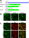
Role of TMH1 in the targeting and retention of C22DES in the ER membrane. a Schematic representation of the GFP fusion constructs generated to study the role of TMH and MCR1 in the targeting and retention of C22DES in the ER. Grey boxes indicate the transmembrane helix (TMH), orange boxes correspond to the MCR1 motif and green boxes correspond to the GFP protein. The amino acid sequence of TMH and MCR1 are shown below the corresponding regions. b Confocal optical sections showing the GFP fluorescence pattern of N. benthamiana cells transiently expressing TMH + MCR1-GFP, TMH-GFP, and MCR1-GFP. The arrow indicates the cell nucleus (n). c Close-up view of the fluorescence pattern of TMH-GFP and MCR1-GFP (left), T3RE (middle) and the corresponding merged images (right)
Fig. 5
Fig. 5
Targeting and retention of the C22DES globular domain in the ER. a Confocal optical sections showing the fluorescence of C22DESΔ2–27-RFP (left) and TMH-GFP (middle) transiently expressed in N. benthamiana leaves. Merged images are shown on the right. b FRAP curves representing the fluorescence recovery rates of C22DESΔ2–27-RFP, BRL3-GFP, and GFP. Fluorescence recovery curves represent the best fits from normalized datasets of at least 6 independently bleached points spots. (C) Cropped images of immunoblot analysis of soluble (S) and membrane (M) cell fractions from N. benthamiana leaves transiently expressing BRL3-GFP (≈153 kDa) and GFP (≈26.8 kDa) as membrane bound and soluble control proteins, respectively. Full-length blots are presented in Additional file 6: Fig. S4. c Cropped images from immunoblot analysis of soluble (S) and membrane (M) cell fractions from N. benthamiana leaves transiently expressing C22DESΔ2–27-GFP (≈84.2 kDa), TMH-GFP (≈34 kDa), and C22DES-GFP (≈87.2 kDa). Full-length blots are shown in Additional file 7: Fig. S5
Fig. 6
Fig. 6
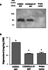
Test of C22DESΔ2–27 enzymatic activity in vivo (a) Cropped image from immunoblot analysis of C22DES-GFP (≈87.24 kDa) and C22DESΔ2–27-GFP (≈84.22 kDa) of agroinfiltrated N. benthamiana leaves. Full-length blot is presented in Additional file 8: Fig. S6. b Stigmasterol levels in total sterol fractions of N. benthamiana leaves expressing C22DES-GFP and C22DESΔ2–27-GFP. Values are mean values ± SD of three technical replicates (n = 3). Lowercase letters indicate significant differences among mean values relative to those in leaf samples expressing the empty vector (one-way ANOVA with Dunnett’s multiple comparisons test)
Fig. 7
Fig. 7
Multiple sequence alignment of the N-terminal region of plant C22DES. The sequence alignment of the N-terminal region of C22DES from the 27 plant species listed in Additional file 2: Table S1 is shown. Amino acid residues are numbered on the right. Asterisks denote residues conserved in all sequences. Colons indicate conservation between amino acid groups of strongly similar properties whereas periods indicate conservation between amino acid groups of weakly similar properties. Hyphens indicate gaps introduced to optimize the alignment. The tomato TMH sequence is highlighted in blue; prolines (P) are shown in red, and serine (S) and threonine (T) residues are shown in magenta. The CRAC1 motif (including the conserved Q27 and Y30 residues) and the MCR1 sequence are also shown. The branched-chain amino-acids [leucine (L), valine (V) and isoleucine (I)] in CRAC1 are shown in green, tyrosine (Y) in cyan and the dibasic residues [arginine (R) and lysine (K)] in yellow. Soly, Solanum lycopersicum; Klni, Klebsormidium nitens; Semo, Selaginella moellendorffii; Pisy, Pinus sylvestris; Sppo, Spirodela polyrhiza; Orsa, Oryza sativa; Deca, Dendrobium catenatum; Phda, Phoenix dactylifera; Anco, Ananas comosus; Amtr, Amborella trichopoda; Bevu, Beta vulgaris; Kafe, Kalanchoe fedtschenkoi; Potr, Populus trichocarpa; Cicl, Citrus clementina; Gora, Gossypium raimondii; Arth, Arabidopsis thaliana; Frve, Fragaria vesca; Ergu, Erythranthe guttata; Cusa, Cucumis sativus; Glma, Glycine max; Paso, Papaver somniferum; Jure, Juglans regia; Nenu, Nelumbo nucifera; Daca, Daucus carota; Cyca, Cynara cardunculus; Vivi, Vitis vinifera; Eugr, Eucalyptus grandis

Similar articles

Cited by

References

    1. Demel RA, De Kruyff B. The function of sterols in membranes. Biochem Biophys Acta. 1976;457:109–132. - PubMed
    1. Hartmann-Bouillon M-A, Benveniste P. Sterol biosynthetic capability of purified membrane fractions from maize coleoptiles. Phytochemistry. 1978;17:1037–1042. doi: 10.1016/S0031-9422(00)94275-4. - DOI
    1. Hartmann M-A. Plant sterols and the membrane environment. Trends Plant Sci. 1998;3(5):170–175. doi: 10.1016/S1360-1385(98)01233-3. - DOI
    1. Ferrer A, Altabella T, Arró M, Boronat A. Emerging roles for conjugated sterols in plants. Prog Lipid Res. 2017;67:27–37. doi: 10.1016/j.plipres.2017.06.002. - DOI - PubMed
    1. Boutté Y, Grebe M. Cellular processes relying on sterol function in plants. Curr Opin Plant Biol. 2009;12:705–713. doi: 10.1016/j.pbi.2009.09.013. - DOI - PubMed

MeSH terms