Case Reports
. 2021 Mar 17;21(1):119.
doi: 10.1186/s12883-021-02144-5.

Venous hypertension caused by a meningioma involving the sigmoid sinus: case report

Affiliations
Case Reports

Venous hypertension caused by a meningioma involving the sigmoid sinus: case report

Koichiro Sumi et al. BMC Neurol. .

Abstract

Background: Intracranial venous hypertension has been associated with a few cases of meningioma secondary to compression of the venous sinus. This is the rare case of small meningioma involving the sigmoid sinus leading to intracranial venous hypertension mimicking venous thrombosis.

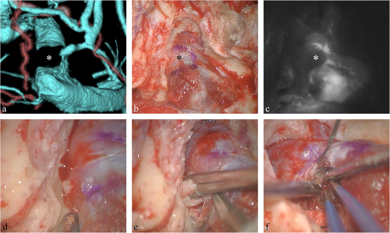
Case presentation: A 39-year-old woman suffered visual dysfunction due to bilateral papilledema. Noncontrast head computed tomography (CT) showed no intracranial space-occupying lesions or hydrocephalus. Cerebrospinal fluid examination revealed high opening pressure. Various image inspections such as three-dimensional CT angiography, magnetic resonance imaging, and cerebral angiography demonstrated a small 2.5-cm lesion causing subtotal occlusion of the dominant right sigmoid sinus. No improvement of clinical manifestations was observed after medical treatment for 6 months, so right presigmoid craniectomy was performed. Operative findings revealed that the tumor was located predominantly involving the sigmoid sinus. The pathological diagnosis was fibrous meningioma. Postoperative fundoscopic examination showed improvement of bilateral papilledema.

Conclusions: We treated a patient presenting with intracranial hypertension due to a small meningioma involving the sigmoid sinus. This unusual case suggests that early surgical strategies should be undertaken to relieve the sinus obstruction.

Keywords: Case report; Meningioma; Sigmoid sinus; Venous hypertension.


Conflict of interest statement

All authors have no affiliations with or involvement in any organization or entity with any financial interest, or non-financial interest, in the subject matter or materials discussed in this case report.

Figures

Fig. 1
Fig. 1
a Preoperative computed tomography (CT) scan revealing no intracranial space-occupying lesions or hydrocephalus except an asymptomatic arachnoid cyst of the left middle fossa. bc Preoperative magnetic resonance images showing the lesion in the right sigmoid sinus (arrowhead) appearing as isointense on T1-weighted image (b), and with homogeneous enhancement following intravenous administration of gadolinium (c). No obvious dural tail sign is present. d Sagittal gadolinium-enhanced T1-weighted images revealing the mass lesion (arrowhead) located under the transverse sinus (arrow). Mass lesion causes severe luminal narrowing. e Conventional angiogram demonstrating the dominant right transverse sinus with hypoplastic left transverse sinus, and subtotal occlusion of the dominant right sigmoid sinus in the venous phase, with antegrade right transverse flow and poor collateral flow indicating this flow is dependent even in subtotal occlusion. f Conventional angiogram, venous phase, showing the mass lesion apparently located at the inner sinus wall mimicking venous thrombosis
Fig. 2
Fig. 2
a Operative CT venogram demonstrating subtotal occlusion of the right sigmoid sinus, with severe luminal narrowing on the posterior side. The emissary vein was found downstream of the severe stenosis. The tumor (asterisk) is located inside the sigmoid sinus. b Right presigmoid craniectomy was performed. After drilling the petrosal bones, very high pressure was found in the transverse and sigmoid sinuses. c Intraoperative indocyanine green fluorescence angiogram revealing a mass lesion as a blood flow defect. The mass was located inside the sigmoid sinus. df The tumor was observed after retraction of the dura and sinus, without dura or sinus wall incision. The tumor was extruded out spontaneously, because of the high pressure in the sinus. The extra sinus part of the tumor was removed first, and then the tumor was followed into the sinus. The tumor was located predominantly involving the sigmoid sinus
Fig. 3
Fig. 3
a Postoperative gadolinium-enhanced T1-weighted magnetic resonance images showing removal of the tumor (arrowhead). b Postoperative cerebral angiography showed patency of the left transverse and sigmoid sinuses showing removal of the tumor (arrowhead)

References

    1. Alvernia JE, Sindou MP. Torcular, transverse, and sigmoid sinus meningiomas. In: Lee JH, editor. Meningiomas. London: Springer; 2009. pp. 473–483.
    1. Taki N, Wein RO, Bedi H, Heilman CB. Extracranial intraluminal extension of atypical meningioma within the internal jugular vein. Case Rep Otolaryngol. 2013;2013:875607. doi: 10.1155/2013/875607. - DOI - PMC - PubMed
    1. Yamashiro K, Hasegawa M, Higashiguchi S, Kato H, Hirose Y. Intravenous sinus meningioma with intraluminal extension to the internal jugular vein: case report and review of the literature. Br J Neurosurg. 2020:1–6. 10.1080/02688697.2020.1777258. - PubMed
    1. Sindou M. Meningiomas invading the sagittal or transverse sinuses, resection with venous reconstruction. J Clin Neurosci. 2001;8(Suppl 1):8–11. doi: 10.1054/jocn.2001.0868. - DOI - PubMed
    1. Szitkar B. A meningioma exclusively located inside the superior sagittal sinus responsible for intracranial hypertension. AJNR Am J Neuroradiol. 2010;31(6):E57–E58. doi: 10.3174/ajnr.A2130. - DOI - PMC - PubMed

Publication types

LinkOut - more resources