

A comparison of standard and high dose adenosine protocols in routine vasodilator stress cardiovascular magnetic resonance: dosage affects hyperaemic myocardial blood flow in patients with severe left ventricular systolic impairment
- PMID: 33731141
- PMCID: PMC7971951
- DOI: 10.1186/s12968-021-00714-7
A comparison of standard and high dose adenosine protocols in routine vasodilator stress cardiovascular magnetic resonance: dosage affects hyperaemic myocardial blood flow in patients with severe left ventricular systolic impairment
Abstract
Background: Adenosine stress perfusion cardiovascular magnetic resonance (CMR) is commonly used in the assessment of patients with suspected ischaemia. Accepted protocols recommend administration of adenosine at a dose of 140 µg/kg/min increased up to 210 µg/kg/min if required. Conventionally, adequate stress has been assessed using change in heart rate, however, recent studies have suggested that these peripheral measurements may not reflect hyperaemia and can be blunted, in particular, in patients with heart failure. This study looked to compare stress myocardial blood flow (MBF) and haemodynamic response with different dosing regimens of adenosine during stress perfusion CMR in patients and healthy controls.
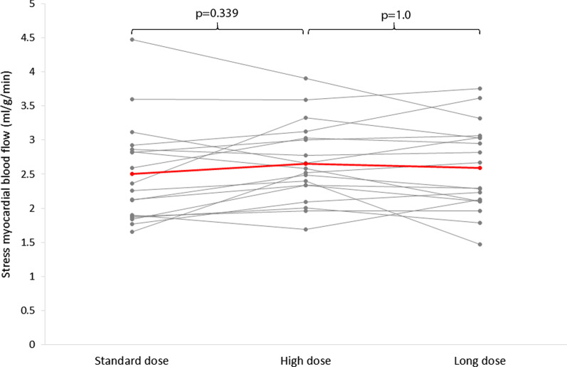
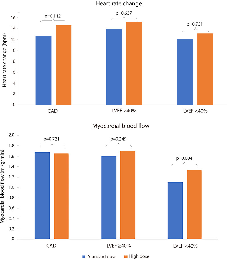
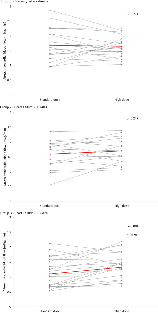
Methods: 20 healthy adult subjects were recruited as controls to compare 3 adenosine perfusion protocols: standard dose (140 µg/kg/min for 4 min), high dose (210 µg/kg/min for 4 min) and long dose (140 µg/kg/min for 8 min). 60 patients with either known or suspected coronary artery disease (CAD) or with heart failure and different degrees of left ventricular (LV) dysfunction underwent adenosine stress with standard and high dose adenosine within the same scan. All studies were carried out on a 3 T CMR scanner. Quantitative global myocardial perfusion and haemodynamic response were compared between doses.
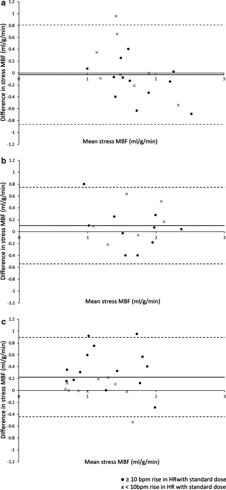
Results: In healthy controls, no significant difference was seen in stress MBF between the 3 protocols. In patients with known or suspected CAD, and those with heart failure and mild systolic impairment (LV ejection fraction (LVEF) ≥ 40%) no significant difference was seen in stress MBF between standard and high dose adenosine. In those with LVEF < 40%, there was a significantly higher stress MBF following high dose adenosine compared to standard dose (1.33 ± 0.46 vs 1.10 ± 0.47 ml/g/min, p = 0.004). Non-responders to standard dose adenosine (defined by an increase in heart rate (HR) < 10 bpm) had a significantly higher stress HR following high dose (75 ± 12 vs 70 ± 14 bpm, p = 0.034), but showed no significant difference in stress MBF.
Conclusions: Increasing adenosine dose from 140 to 210 µg/kg/min leads to increased stress MBF in patients with significantly impaired LV systolic function. Adenosine dose in clinical perfusion assessment may need to be increased in these patients.
Keywords: Adenosine stress; Heart failure; Myocardial blood flow; Perfusion.
Conflict of interest statement
The authors declare that they have no competing interests.
Figures
References
-
- Greenwood JP, Maredia N, Younger JF, Brown JM, Nixon J, Everett CC, et al. Cardiovascular magnetic resonance and single-photon emission computed tomography for diagnosis of coronary heart disease (CE-MARC): a prospective trial. Lancet. 2012;379(379):453–460. doi: 10.1016/S0140-6736(11)61335-4. - DOI - PMC - PubMed
-
- Greenwood JP, Ripley DP, Berry C, McCann GP, Plein S, Bucciarelli-Ducci C, et al. Effect of care guided by cardiovascular magnetic resonance, myocardial perfusion scintigraphy, or NICE guidelines on subsequent unnecessary angiography rates : the CE-MARC 2 randomized clinical trial. JAMA. 2016;316(10):1051–1060. doi: 10.1001/jama.2016.12680. - DOI - PubMed
Publication types
MeSH terms
Substances
Grants and funding
LinkOut - more resources
Full Text Sources
Other Literature Sources
Miscellaneous
