. 2021 Mar 18;23(1):28.
doi: 10.1186/s12968-021-00719-2.

Usefulness of TI-scout images in the assessment of late gadolinium enhancement in children

Affiliations

Usefulness of TI-scout images in the assessment of late gadolinium enhancement in children

Badr Bannan et al. J Cardiovasc Magn Reson. .

Abstract

Background: Cardiovascular magnetic resonance (CMR) late gadolinium enhancement (LGE) requires identification of the normal myocardial nulling time using inversion time (TI)-scout imaging sequence. Although TI-scout images are not primarily used for myocardial assessment, they provide information regarding different signal recovery patterns of normal and abnormal myocardium facilitating identification of LGE in instances where standard LGE images alone are not diagnostic. We aimed to assess the diagnostic performance of TI-scout as compared to that of standard LGE images.

Methods: CMR studies with LGE imaging in 519 patients (345 males, 1-17 years) were reviewed to assess the diagnostic performance of LGE imaging in terms of the location of LGE and the pathologic entities. The diagnostic performance of the TI-scout and standard LGE imaging was classified into four categories: (1) equally diagnostic, (2) TI-scout superior to standard LGE, (3) standard LGE superior to TI-scout, and (4) complementary, by the consensus of the two observers.

Results: The study cohort consisted of 440 patients with negative LGE and 79 with evidence for LGE. For a negative diagnosis of LGE, TI-scout and standard LGE images were equally diagnostic in 75% of the cases and were complementary in 12%. For patients with LGE, TI-scout images were superior to standard LGE images in 52% of the cases and were complementary in 19%. The diagnostic performance of TI-scout images was superior to that of standard LGE images in all locations. TI-scout images were superior to standard LGE images in 11 of 12 (92%) cases with LGE involving the papillary muscles, in 7 /12 (58%) cases with subendocardial LGE, and in 4/7 (57%) cases with transmural LGE. TI-scout images were particularly useful assessing the presence and extent of LGE in hypertrophic cardiomyopathy (HCM). TI-scout was superior to standard LGE in 6/10 (60%) and was complementary in 3/10 (30%) of the positive cases with HCM.

Conclusions: TI-scout images enhance the diagnostic performance of LGE imaging in children.

Keywords: Fibrosis; Late gadolinium enhancement; Look-Locker; Magnetic resonance; Myocardium; TI-scout.


Conflict of interest statement

The authors declare that they have no competing interests.

Figures
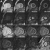
Fig. 1
Fig. 1
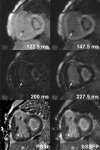
TI-scout images showing different signal intensity recovery after inversion pulse in normal (a) and abnormal (b) myocardium. In images obtained from a patient with aortic stenosis (b), there is obvious endocardial/subendocardial fibrosis in all TI-scout images with changing signal intensity. Note that late gadolinium enhancement (LGE) is barely identifiable in phase sensitive inversion recovery (PSIR) image in right lower corner
Fig. 2
Fig. 2
Late gadolinium enhancement (LGE) in a 13-year old with Duchenne muscular dystrophy. A similar extent of LGE involving subepicaridal/mid-myocardial layer of the inferolateral and lateral wall and papillary muscles of the left ventricle is seen in both TI (inversion time)-scout and standard LGE images, playing complementary roles in the diagnosis of LGE. Phase sensitive inversion recovery (PSIR) and balanced steady-state free precession (bSSFP) standard LGE images were obtained at TI of 330 ms
Fig. 3
Fig. 3
Late gadolinium enhancement (LGE) in a 6-month old with critical aortic and mitral stenosis. The presence and extent of LGE involving endocardium and subendocardium is appreciated better in TI (inversion time)-scout images than in phase sensitive inversion recovery (PSIR) and balanced steady state free precession (bSSFP) images. LGE of a papillary muscle (arrows) in the right ventricle is identifiable only in TI-scout images. Phase sensitive inversion recovery (PSIR) and balanced steady-state free precession (bSSFP) standard LGE images were obtained at TI of 280 ms
Fig. 4
Fig. 4
Focal subendocardial fibrosis in a 14-year old with concentric hypertrophic cardiomyopathy. A focal subendocardial fibrosis is well appreciated in TI (inversion time)-scout images. In phase sensitive inversion recovery (PSIR) and balanced steady-state free precession (bSSFP) images, high signal intensity involving the same region may represent either true late gadolinium enhancement (LGE) or blood pool in a myocardial crypt. Phase sensitive inversion recovery (PSIR) and steady-state free precession (SSFP) standard LGE images were obtained at TI of 310 ms
Fig. 5
Fig. 5
Heterogeneous mild late gadolinium enhancement (LGE) in a 16-year old with with hypertrophic cardiomyopathy. Heterogeneity of the signal intensity of the hypertrophic myocardium can be confidently appreciated only in TI (inversion time)-scout images. PSIR and bSSFP standard LGE images were obtained at TI of 280 ms

References

    1. Kellman P, Arai AE. Cardiac imaging techniques for physicians: late enhancement. J Magn Reson Imaging. 2012;36:529–42. doi: 10.1002/jmri.23605. - DOI - PMC - PubMed
    1. Amano Y, Takayama M, Kumita S. Contrast-enhanced myocardial T1-weighted scout (Look-Locker) imaging for the detection of myocardial damages in hypertrophic cardiomyopathy. J Magn Reson Imaging. 2009;30:778–84. doi: 10.1002/jmri.21921. - DOI - PubMed
    1. Pandey T, Jambhekar K, Shaikh R, Lensing S, Viswamitra S. Utility of the inversion scout sequence (TI scout) in diagnosing myocardial amyloid infiltration. Int J Cardiovasc Imaging. 2013;29:103–12. doi: 10.1007/s10554-012-0042-4. - DOI - PMC - PubMed
    1. Kellman P, Xue H, Olivieri LJ, Cross RR, Grant EK, Fontana M, et al. Dark blood late enhancement imaging. J Cardiovasc Magn Reson. 2016;18:77. doi: 10.1186/s12968-016-0297-3. - DOI - PMC - PubMed
    1. Francis R, Kellman P, Kotecha T, Baggiano A, Norrington K, Martinez-Naharro A, et al. Prospective comparison of novel dark blood late gadolinium enhancement with conventional bright blood imaging for the detection of scar. J Cardiovasc Magn Reson. 2017;19:91. doi: 10.1186/s12968-017-0407-x. - DOI - PMC - PubMed