. 2021 Apr 15;39(16):2280-2287.
doi: 10.1016/j.vaccine.2021.03.006. Epub 2021 Mar 5.

Intranasal administration of a recombinant RBD vaccine induced protective immunity against SARS-CoV-2 in mouse

Affiliations

Intranasal administration of a recombinant RBD vaccine induced protective immunity against SARS-CoV-2 in mouse

Yingying Du et al. Vaccine. .

Abstract

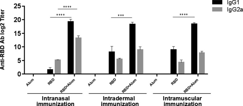
The emergence of the global Severe Acute Respiratory Syndrome Coronavirus-2 (SARS-CoV-2) pandemic underscores the importance of the rapid development of a non-invasive vaccine that can be easily administered. A vaccine administered by nasal delivery is endowed with such characteristics against respiratory viruses. In this study, we generated a recombinant SARS-CoV-2 receptor-binding domain (RBD)-based subunit vaccine. Mice were immunized via intranasal inoculation, microneedle-intradermal injection, or intramuscular injection, after which the RBD-specific immune responses were compared. Results showed that when administrated intranasally, the vaccine elicited a robust systemic humoral immunity with high titers of IgG antibodies and neutralizing antibodies as well as a significant mucosal immunity. Besides, antigen-specific T cell responses were also analyzed. These results indicated that the non-invasive intranasal administration should be explored for the future SARS-CoV-2 vaccine design.

Keywords: Intranasal administration; Mucosal immunity; Neutralizing antibody; SARS-CoV-2.


Conflict of interest statement

Declaration of Competing Interest The authors declare that they have no known competing financial interests or personal relationships that could have appeared to influence the work reported in this paper.

Figures

Fig. 1
Fig. 1
Characterization of recombinant RBD and immunization schedule of mice. (A) Analysis of purified RBD. Rabbit anti-RBD polyclonal antibody was used for RBD detection. Left panel: SDS-PAGE of purified RBD. Right panel: Western blot of purified RBD. (B) Schematic of the vaccination schedule of mice. They were immunized three times at intervals of 3 weeks through intranasal immunization (i.n.), intradermal immunization (i.d.), or intramuscular immunization (i.m.). Blood samples were collected before each boost immunization and before sacrifice. Vaginal washes were collected for 3 consecutive days before sacrifice. Samples of spleens, nasal washes, bronchoalveolar lavage fluids, and intestines were collected at the time of sacrifice.
Fig. 2
Fig. 2
SARS-CoV-2 RBD-specific IgG antibody response in mice after each immunization, as measured by ELISA. i.n.: intranasal immunization, i.d.: intradermal immunization, i.m.: intramuscular immunization. Results are expressed as the mean value ± standard error of the mean (SEM) of seven mice in each group. **0.001 < P < 0.01.
Fig. 3
Fig. 3
SARS-CoV-2 RBD-specific IgG1 and IgG2a titers in mice 3 weeks after the last immunization. Results are expressed as the mean value ± standard error of the mean (SEM) of six mice in each group. ***0.0001 < P < 0.001; ****P < 0.0001.
Fig. 4
Fig. 4
SARS-CoV-2 RBD-specific secretory IgA titers in (A) nasal washes, (B) bronchoalveolar lavage fluids, (C) vaginal washes, and (D) intestines of mice 3 weeks after the last immunization. Results are expressed as the mean value ± standard error of the mean (SEM) of six mice in each group.
Fig. 5
Fig. 5
ELISpot detection of IFN-γ producing cells (A, C, and E) and IL-4 producing cells (B, D, and F) in immunized mice. i.n.: intranasal immunization, i.d.: intradermal immunization, i.m.: intramuscular immunization. Results are expressed as spot-forming cells (SFCs) per 106 spleen cells.
Fig. 6
Fig. 6
Cell–cell fusion inhibition assay. (A) Principle of S protein-mediated cell–cell fusion assay. (B) Serum samples in each group were tested in duplicate. Data are presented as relative light units (RLUs) %.

Similar articles

Cited by

References

    1. Rogers TF, Zhao F, Huang D, Beutler N, Burns A, He WT, et al. Isolation of potent SARS-CoV-2 neutralizing antibodies and protection from disease in a small animal model. Science (New York, N.Y.); 2020; eabc7520. Advance online publication. - PMC - PubMed
    1. Alturki S.O., Alturki S.O., Connors J., Cusimano G., Kutzler M.A., Izmirly A.M., et al. The 2020 pandemic: current SARS-CoV-2 vaccine development. Front Immunol. 2020 Aug 19;11:1880. doi: 10.3389/fimmu.2020.01880. PMID: 32973779; PMCID: PMC7466534. - DOI - PMC - PubMed
    1. Du L., Zhao G., Li L., He Y., Zhou Y., Zheng B.J., Iang S. Antigenicity and immunogenicity of SARS-CoV S protein receptor-binding domain stably expressed in CHO cells. Biochem Biophys Res Commun. 2009;384(4):486–490. - PMC - PubMed
    1. Du L., Zhao G., Chan C.C., Sun S., Chen M., Liu Z., et al. Recombinant receptor-binding domain of SARS-CoV spike protein expressed in mammalian, insect and E. coli cells elicits potent neutralizing antibody and protective immunity. Virology. 2009;393(1):144–150. - PMC - PubMed
    1. He Y., Zhou Y., Liu S., Kou Z., Li W., Farzan M., et al. Receptor-binding domain of SARS-CoV spike protein induces highly potent neutralizing antibodies: implication for developing subunit vaccine. Biochem Biophys Res Commun. 2004;324(2):773–781. - PMC - PubMed

Publication types

MeSH terms