. 2021 Mar 17;7(12):eabe2470.
doi: 10.1126/sciadv.abe2470. Print 2021 Mar.

GREB1: An evolutionarily conserved protein with a glycosyltransferase domain links ERα glycosylation and stability to cancer

Affiliations

GREB1: An evolutionarily conserved protein with a glycosyltransferase domain links ERα glycosylation and stability to cancer

Eun Myoung Shin et al. Sci Adv. .

Abstract

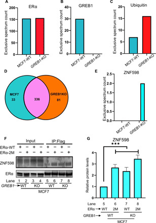
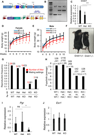
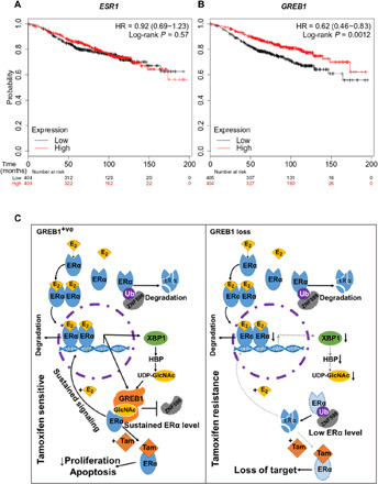
What covalent modifications control the temporal ubiquitination of ERα and hence the duration of its transcriptional activity remain poorly understood. We show that GREB1, an ERα-inducible enzyme, catalyzes O-GlcNAcylation of ERα at residues T553/S554, which stabilizes ERα protein by inhibiting association with the ubiquitin ligase ZNF598. Loss of GREB1-mediated glycosylation of ERα results in reduced cellular ERα levels and insensitivity to estrogen. Higher GREB1 expression in ERα+ve breast cancer is associated with greater survival in response to tamoxifen, an ERα agonist. Mice lacking Greb1 exhibit growth and fertility defects reminiscent of phenotypes in ERα-null mice. In summary, this study identifies GREB1, a protein with an evolutionarily conserved domain related to DNA-modifying glycosyltransferases of bacteriophages and kinetoplastids, as the first inducible and the only other (apart from OGT) O-GlcNAc glycosyltransferase in mammalian cytoplasm and ERα as its first substrate.


Figures

Fig. 1
Fig. 1. GREB1 regulates ERα signaling through stabilization of ERα.
(A) Schematic diagram of predicted GREB1 domains (17). (B) Heatmap illustrating the top 10 up-regulated and down-regulated genes from RNA sequencing of MCF7-WT cells with or without estradiol (E2) stimulation. (C) MCF7-WT and GREB1-KO cells were treated with E2 and analyzed by qPCR. n = 3; ns, not significant, *P < 0.05, ***P < 0.001, and ****P < 0.0001 by two-way analysis of variance (ANOVA) with Holm-Sidak’s multiple comparisons test. (D) ERα recruitment at indicated promoters in MCF7-WT and GREB1-KO cells were analyzed by ChIP followed by qPCR. n = 2; ***P < 0.001 and ****P < 0.0001 by one-way ANOVA with Holm-Sidak’s multiple comparisons test. (E) Schematic illustration of the GREB1 construct used for immunofluorescence. (F) GREB1 localization was analyzed by immunofluorescence microscopy in HeLa cells expressing the tagged GREB1 construct. Nuclei were stained with DAPI. (G) In MCF7-WT and GREB1-KO cells, levels of indicated proteins were analyzed by Western blot. (H and I) After E2 treatment in MCF7-WT and GREB1-KO cells, protein levels (H) and ESR1 mRNA expression (I) were analyzed. n = 2; by ANOVA two-way test. (J) MCF7-WT cells were transduced with empty vector (Vec) or GREB1 construct and subsequently treated with CHX. (K) GREB1-KO cells were transduced with empty Vec or GREB1 construct and subsequently treated with CHX.
Fig. 2
Fig. 2. GREB1 GT activity is important in stabilizing ERα.
(A) FLAG pulldown was analyzed by Western blot in MCF7-WT or GREB1-KO cells expressing FLAG-ERα. (B) Catalytically dead mutant GREB1 (GT-Mut) was generated by mutating indicated residues in GREB1’s GT domain. (C) The growth of GREB1-KO cells transduced with Vec, GT-WT, or GT-Mut was assessed by colony formation assay with crystal violet staining. Photo credit: Sultan Abda Neja, Institute of Cell and Molecular Biology, A*STAR. (D and E) GREB1-KO cells transduced with GT-WT or GT-Mut were subcutaneously injected to the flanks of the same NOD/SCID mice, each cell line on one side. The resulting tumors in these mice were harvested (D) and weighted (E). n = 8; ****P < 0.0001 by unpaired t test. Photo credit: Anandhkumar Raju, Institute of Molecular and Cell Biology, A*STAR. (F) GREB1-KO cells transduced with Vec, GT-WT, or GT-Mut. (G) ERα, GT-WT, and GT-Mut were individually purified from 293T. In vitro assay using a UDP-Glo Glycosyltransferase Assay kit was performed as indicated. n = 3. (H) S. cerevisiae yeast was transfected with either Vec or FLAG-conjugated GREB1 construct.
Fig. 3
Fig. 3. GREB1 glycosylates ERα on T553 and S554.
(A) HEK293T cells were transfected with FLAG-ERα together with either Vec, GT-WT, or GT-Mut. (B) FLAG pulldown was performed in HEK293T cells described in (A). Total protein eluates were analyzed by Coomassie blue staining. (C to E) O-GlcNAcylation of gel-extracted ERα from the corresponding band from (B) was labeled using BEMAD [β-elimination and Michael addition with dithiothreitol (DTT)] methodology and analyzed by LC-MS/MS. Spectra of ERα tryptic peptide LHAPTSR with modifications at T553 (C) and S554 (D) and with no modification (E) are illustrated. (F) Quantification of the fraction of peptides that were glycosylated at T553 and S554 in the indicated samples. n = 2; *P < 0.05 by one-way ANOVA with Holm-Sidak’s multiple comparisons test. (G) Quantification of the fraction of peptides that were glycosylated at S10 in the indicated samples. n = 2; by one-way ANOVA with Holm-Sidak’s multiple comparisons test.
Fig. 4
Fig. 4. Glycosylation on T553 and S554 is important for GREB1 stabilization of ERα.
(A) Schematic of ESR1 WT construct (ERα-WT) or mutated construct (ERα-2M). (B) ERα-WT and ERα-2M expressed by HEK293T (left) were analyzed for DNA binding capability by electrophoretic mobility shift assay (EMSA) (right panel). (C and D) MCF7-WT cells (C) and GREB1-KO cells (D) were transduced with Vec, ERα-WT, or ERα-2M construct and subsequently treated with CHX. (E) HEK293T cells were transfected with GREB1 alongside either ERα-WT or ERα-2M constructs, and FLAG pulldown was performed. Two replicates (Rep.) were analyzed together on Western blot. (F and G) MCF7 cells were transfected with either control siRNA (siControl) or XBP1 siRNA (siXBP1). Cells were treated with or without E2 for 24 hours and subsequently subjected to a CHX chase assay. Levels of indicated proteins were analyzed by Western blot (F), and the amount of the remaining ERα after CHX treatment was analyzed by densitometry quantification (G). n = 3, **P < 0.01 by t test.
Fig. 5
Fig. 5. GREB1 stabilizes ERα by limiting binding of the ubiquitin ligase ZNF598.
(A to E) MCF7-WT or GREB1-KO cells were transduced with FLAG-ERα, and FLAG pulldown was performed. Eluates were analyzed by MS. The number of peptide reads for ERα (A) and GREB1 (B) was examined as controls. The number of peptide reads for ubiquitin (C), unique or common interactors (D), and ZNF598 (E) from MCF7-WT and GREB1-KO was illustrated. (F) FLAG pulldown was performed to MCF7-WT or GREB1-KO cells transduced with ERα-WT or ERα-2M. (G) Densitometry analysis of ZNF598 bands from the Western blot in (F) for corresponding lanes. n = 3; ***P < 0.005 by one-way ANOVA.
Fig. 6
Fig. 6. Greb1 KO reduces growth and fertility in female mice.
(A) Schematic presentation of the generation of Greb1-KO mice by inserting a trapping cassette “SA-βgeo-pA” upstream exons 16 and 17 of Greb1 and corresponding allele expressing GREB1-WT when the trapping cassette is removed by Flp-recombinase targeting FRT sites flanking the SA-βgeo-pA cassette. (B) Representative genotyping for WT, Het, and Greb1-KO mice by PCR. (C) Mouse ovaries were harvested from WT, Het, or Greb1-KO mice. mRNA level of Greb1 was evaluated by qRT-PCR. n = 3, ***P < 0.001 by t test. (D) Body weight (in grams) of WT or Greb1-KO female mice. *P < 0.05 and **P < 0.01 by t test. (E) Body weight (in grams) of WT or Greb1-KO male mice. (F) Representative photo image of age-matched WT and Greb1-KO female mice at 25 days. Photo credit: Chia Yi Liu, Institute of Molecular and Cell Biology, A*STAR. (G and H) A total of six rounds of mattings were performed with multiple mating cages, one male and two female mice per mating cage, for each pairing consisting of indicated genotypes. In different mating combinations of WT, Het, and Greb1-KO mice, the frequency of conceptions was calculated as the ratio between the number of litter conceived per number of mating cages (G), and the average number of pups per litter from different mating settings of WT and Greb1-KO mice was calculated (H). (I) Ovary samples from WT, Het, and Greb1-KO mice were collected and analyzed for mouse progesterone receptor (Pgr) expression at mRNA levels. (J) Ovary samples from WT, Het, and Greb1-KO mice were collected and analyzed for mouse estrogen receptor α (Esr1) expression at mRNA levels. n = 3, *P < 0.05 by t test.
Fig. 7
Fig. 7. GREB1 role in tamoxifen response in breast cancer.
(A and B) Kaplan-Meier plots in a cohort of tamoxifen-treated breast cancer patients, plotting tumor’s ESR1 (A) and GREB1 (B) mRNA expression as a function of overall patient survival. (C) Graphical abstracts of GREB1 function in ER+ve breast cancer and drug response. When cells express GREB1, ERα is stabilized by glycosylation and imposes ERα signaling transcription signature, which is vulnerable to tamoxifen, an ERα agonist. For cells that transcriptionally repress GREB1, ERα protein and its transcriptional profile are lost. Because of this loss of target, these cells are resistance to tamoxifen.

References

    1. Lee T. I., Young R. A., Transcriptional regulation and its misregulation in disease. Cell 152, 1237–1251 (2013). - PMC - PubMed
    1. Hayden M. S., Ghosh S., Regulation of NF-κB by TNF family cytokines. Semin. Immunol. 26, 253–266 (2014). - PMC - PubMed
    1. Liu Y., Tavana O., Gu W., p53 modifications: Exquisite decorations of the powerful guardian. J. Mol. Cell Biol. 11, 564–577 (2019). - PMC - PubMed
    1. Hewitt S. C., Korach K. S., Oestrogen receptor knockout mice: Roles for oestrogen receptors alpha and beta in reproductive tissues. Reproduction 125, 143–149 (2003). - PubMed
    1. Tang Z.-R., Zhang R., Lian Z.-X., Deng S.-L., Yu K., Estrogen-receptor expression and function in female reproductive disease. Cells 8, 1123 (2019). - PMC - PubMed

Publication types