. 2021 Apr 28;41(17):3917-3931.
doi: 10.1523/JNEUROSCI.2504-20.2021. Epub 2021 Mar 17.

Reduced Repetition Suppression in Aging is Driven by Tau-Related Hyperactivity in Medial Temporal Lobe

Affiliations

Reduced Repetition Suppression in Aging is Driven by Tau-Related Hyperactivity in Medial Temporal Lobe

Jenna N Adams et al. J Neurosci. .

Abstract

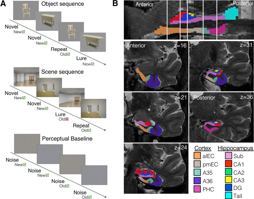
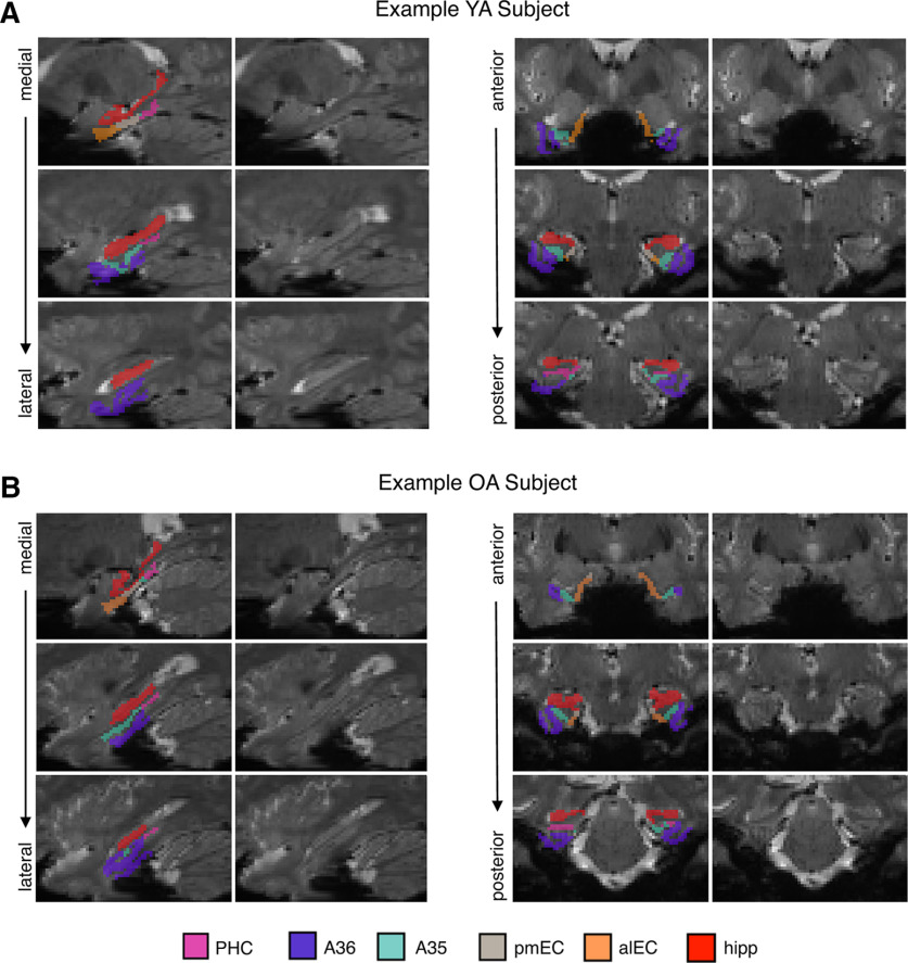
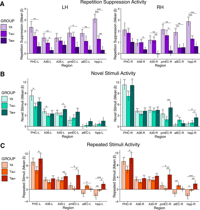
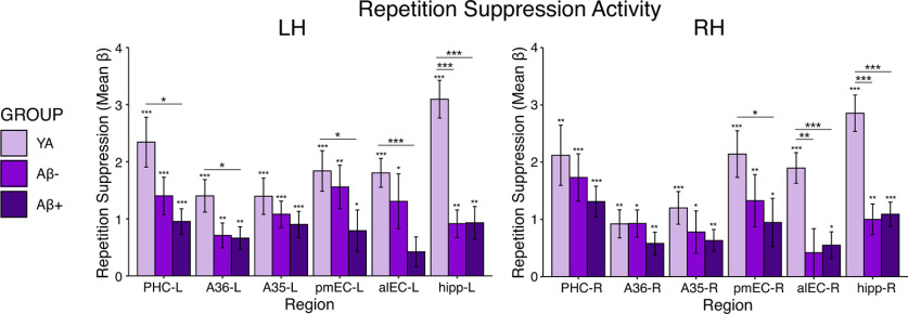
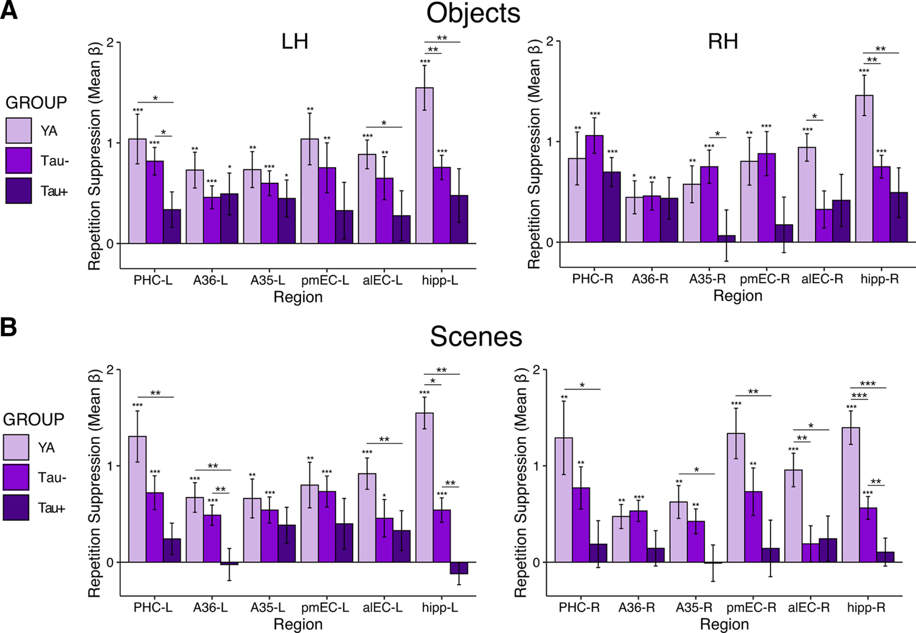
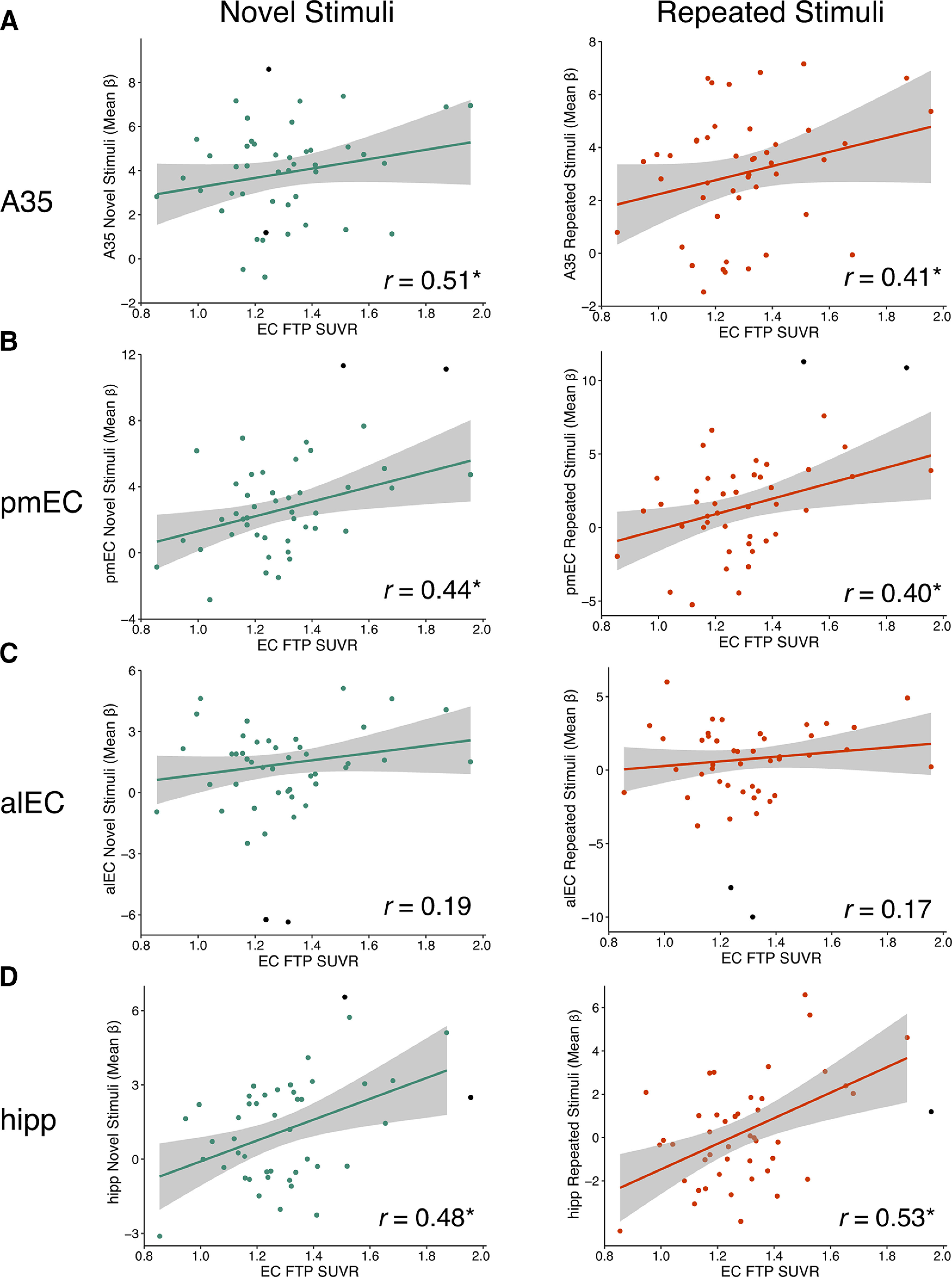
Tau deposition begins in the medial temporal lobe (MTL) in aging and Alzheimer's disease (AD), and MTL neural dysfunction is commonly observed in these groups. However, the association between tau and MTL neural activity has not been fully characterized. We investigated the effects of tau on repetition suppression, the reduction of activity for repeated stimulus presentations compared to novel stimuli. We used task-based functional MRI (fMRI) to assess MTL subregional activity in 21 young adults (YA) and 45 cognitively normal human older adults (OA; total sample: 37 females, 29 males). AD pathology was measured with position emission tomography (PET), using 18F-Flortaucipir for tau and 11C-Pittsburgh compound B (PiB) for amyloid-β (Aβ). The MTL was segmented into six subregions using high-resolution structural images. We compared the effects of low tau pathology, restricted to entorhinal cortex and hippocampus (Tau- OA), to high tau pathology, also occurring in temporal and limbic regions (Tau+ OA). Low levels of tau (Tau- OA vs YA) were associated with reduced repetition suppression activity specifically in anterolateral entorhinal cortex (alEC) and hippocampus, the first regions to accumulate tau. High tau pathology (Tau+ vs Tau- OA) was associated with widespread reductions in repetition suppression across MTL. Further analyses indicated that reduced repetition suppression was driven by hyperactivity to repeated stimuli, rather than decreased activity to novel stimuli. Increased activation was associated with entorhinal tau, but not Aβ. These findings reveal a link between tau deposition and neural dysfunction in MTL, in which tau-related hyperactivity prevents deactivation to repeated stimuli, leading to reduced repetition suppression.SIGNIFICANCE STATEMENT Abnormal neural activity occurs in the medial temporal lobe (MTL) in aging and Alzheimer's disease (AD). Because tau pathology first deposits in the MTL in aging, this altered activity may be due to local tau pathology, and distinct MTL subregions may be differentially vulnerable. We demonstrate that in older adults (OAs) with low tau pathology, there are focal alterations in activity in MTL subregions that first develop tau pathology, while OAs with high tau pathology have aberrant activity throughout MTL. Tau was associated with hyperactivity to repeated stimulus presentations, leading to reduced repetition suppression, the discrimination between novel and repeated stimuli. Our data suggest that tau deposition is related to abnormal activity in MTL before the onset of cognitive decline.

Keywords: Alzheimer's disease; PET; aging; fMRI; repetition suppression; tau.


Figures

Figure 1.
Figure 1.
Visual representation of the fMRI task and MTL segmentation. A, OA and YA subjects performed a mnemonic discrimination task during fMRI acquisition. The first two stimuli of each block (either object or scene sequences) are novel stimuli, while the second two stimuli are either repeated stimuli or highly similar lure stimuli. Subjects are instructed to respond old or new to each stimulus. A perceptual baseline of scrambled noise images was shown at the start and end of the task. Repetition suppression was measured by contrasting activity between all novel and all repeated stimuli. B, High-resolution (0.5 × 0.5 × 1.5 mm) T2 images in native space were segmented with ASHS to derive MTL ROIs for analysis. The full entorhinal cortex was manually subdivided into alEC and pmEC subregions using template ROIs inverse warped to native space. Hippocampal subfields were merged into one hippocampal ROI before analyses. Cortical ROIs: anterolateral entorhinal cortex (alEC), posteromedial EC (pmEC), area 35 of perirhinal cortex (A35), area 36 of perirhinal cortex (A36), parahippocampal cortex (PHC); hippocampal subfields: subiculum (Sub), CA1, CA2, CA3, dentate gyrus (DG), hippocampal tail (Tail).
Figure 2.
Figure 2.
Example MTL segmentations and fMRI signal intensity. Representative images from a YA subject (A) and OA subject (B) are shown to visually demonstrate the quality of fMRI signal in each ROI. For each example subject, the MTL segmentation is overlaid on the mean functional image on the left column, and the mean functional image is shown by itself on the right column. Slices are shown sagittally from medial to lateral (left) and coronally from anterior to posterior (right). ROIs: parahippocampal cortex (PHC), area 36 of perirhinal cortex (A36), area 35 of perirhinal cortex (A35), posteromedial EC (pmEC), anterolateral entorhinal cortex (alEC), hippocampus (hipp; all subfields merged). See Extended Data Figure 2-1 for corresponding proportions of voxels retained in each region in YA and OA groups after removing voxels of low signal.
Figure 3.
Figure 3.
Tau-related differences in MTL activation for repetition suppression, novel stimuli, and repeated stimuli. A, Repetition suppression was quantified by contrasting activity between novel and repeated stimuli, and compared across the YA, low tau (Tau– OA), and high tau (Tau+ OA) groups with ANOVAs and follow-up pairwise comparisons. Tau– OA demonstrated a significant reduction in repetition suppression (vs YA) in bilateral hippocampus and alEC-R (independent samples t test, ps < 0.05). Tau+ OA demonstrated additional reductions in repetition suppression across the MTL (vs YA and Tau– OA; ps < 0.05). B, C, To determine whether activity changes to novel and/or repeated stimuli were driving reduced repetition suppression, we next investigated each subcomponent of the contrast separately. B, Activity to novel stimuli was compared to the perceptual baseline condition. There were minimal group differences in activity to novel stimuli, including both increases and decreases in activity. C, Activity to repeated stimuli was compared to the perceptual baseline condition. Hyperactivity in the Tau– OA group (vs YA) was found in bilateral hippocampus and alEC-R, and hyperactivity in the Tau+ OA group (vs Tau– OA and YA) was found throughout the MTL. This hyperactivity to repeated stimuli can be inferred to drive the reduction in repetition suppression seen in A. Significance above each bar reflects one sample t tests within each group, while lines across bars represent significant group differences with independent samples t tests. Error bars represent SEM; ***p < 0.001, **p < 0.01, *p < 0.05.
Figure 4.
Figure 4.
Aβ-related differences in MTL activation for repetition suppression. Repetition suppression activity was compared across the YA, Aβ– OA, and Aβ+ OA groups with ANOVAs and follow-up pairwise comparisons. While significant differences were found between the YA group and each Aβ group, there were no additional significant difference between Aβ– and Aβ+ OA, indicating no effect of Aβ positivity on repetition suppression activity (ps > 0.05). Significance above each bar reflects one sample t tests within each group, while lines across bars represent significant group differences with independent samples t tests. Error bars represent SEM; ***p < 0.001, **p < 0.01, *p < 0.05.
Figure 5.
Figure 5.
Tau-related differences in MTL activation for repetition suppression for object (A) and scene (B) stimuli modeled separately. Repetition suppression was defined as the difference in activity between the novel and repeated stimuli. Patterns of repetition suppression were generally similar for object (A) and scene (B) stimuli, both regarding patterns within each group and contrasts between groups. Significance above each bar reflects one sample t tests within each age group, while lines across bars represent significant group differences with independent samples t tests. Error bars represent SEM; ***p < 0.001, **p < 0.01, *p < 0.05.
Figure 6.
Figure 6.
Increased activation to novel and repeated stimuli is associated with entorhinal tau pathology in OAs. Mean FTP SUVR of native space entorhinal cortex (EC) was extracted and partial volume corrected for each OA subject. To obtain measures of activity, the mean β value for each ROI was extracted for novel and repeated stimuli (compared to perceptual baseline). Robust regressions were performed to reduce the effects of outliers. All correlations controlled for age, sex, and EC thickness (raw data shown). EC FTP SUVR was associated with increased activity to both novel and repeated stimuli in A35 (A), pmEC (B), and hippocampus (D), but not alEC (C); * indicates significant skipped correlations (CI does not include zero). Black data points were identified as bivariate outliers and removed in the skipped correlation.

Similar articles

Cited by

References

    1. Adams JN, Maass A, Harrison TM, Baker SL, Jagust WJ (2019) Cortical tau deposition follows patterns of entorhinal functional connectivity in aging. Elife 8:e49132. 10.7554/eLife.49132 - DOI - PMC - PubMed
    1. Angulo SL, Orman R, Neymotin SA, Liu L, Buitrago L, Cepeda-Prado E, Stefanov D, Lytton WW, Stewart M, Small SA, Duff KE, Moreno H (2017) Tau and amyloid-related pathologies in the entorhinal cortex have divergent effects in the hippocampal circuit. Neurobiol Dis 108:261–276. 10.1016/j.nbd.2017.08.015 - DOI - PubMed
    1. Avants BB, Yushkevich P, Pluta J, Minkoff D, Korczykowski M, Detre J, Gee JC (2010) The optimal template effect in hippocampus studies of diseased populations. Neuroimage 49:2457–2466. 10.1016/j.neuroimage.2009.09.062 - DOI - PMC - PubMed
    1. Baker SL, Maass A, Jagust WJ (2017) Considerations and code for partial volume correcting [18F]-AV-1451 tau PET data. Data Brief 15:648–657. 10.1016/j.dib.2017.10.024 - DOI - PMC - PubMed
    1. Bakker A, Krauss GL, Albert MS, Speck CL, Jones LR, Stark CE, Yassa MA, Bassett SS, Shelton AL, Gallagher M (2012) Reduction of hippocampal hyperactivity improves cognition in amnestic mild cognitive impairment. Neuron 74:467–474. 10.1016/j.neuron.2012.03.023 - DOI - PMC - PubMed

Publication types

MeSH terms