. 2021 May 9;83(5):784-792.
doi: 10.1292/jvms.20-0532. Epub 2021 Mar 18.

Reduced differentiation of intestinal epithelial cells in wasting marmoset syndrome

Affiliations

Reduced differentiation of intestinal epithelial cells in wasting marmoset syndrome

Kimie Niimi et al. J Vet Med Sci. .

Abstract

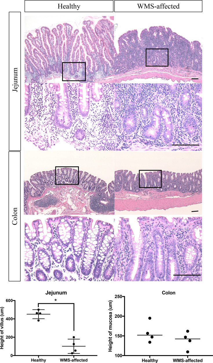
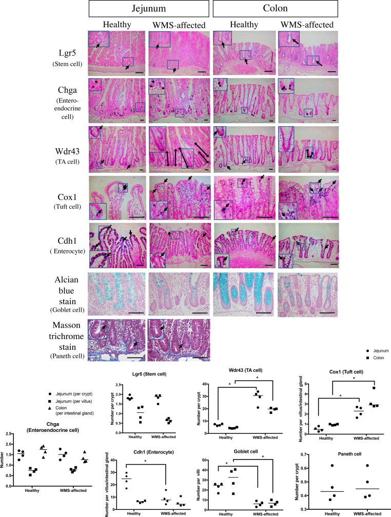
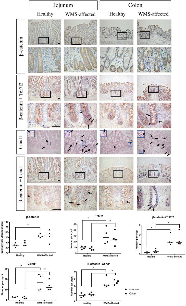
Wasting marmoset syndrome (WMS) is a serious disease in captive common marmoset (Callithrix jacchus) colonies. Because of the high mortality rates, elucidation of the underlying mechanisms is essential. In this study, we compared the histopathology, the number of each epithelial cell in the jejunum and colon, and the expression patterns of some molecular markers between healthy and WMS-affected marmosets. Atrophy of villi in the jejunum and mononuclear cell infiltration in the lamina propria were observed in the intestinal tract of WMS-affected marmosets. Although the numbers of transient amplifying cells and tuft cells were increased, the number of goblet cells was obviously decreased in the jejunum and colon of WMS-affected marmosets compared to healthy marmosets. In addition, the number of enterocytes in the jejunum was decreased in WMS animals. There was no apparent difference in the numbers of stem cells, enteroendocrine cells, or Paneth cells. The expression of β-catenin and Tcf7l2 was increased in WMS, and the co-existence of β-catenin and Tcf7l2/Cyclin D1 was observed around the crypts in WMS-affected marmosets. These findings suggest that cell proliferation continues, but cell differentiation is halted in the intestinal tract due to the enhanced β-catenin/Tcf7l2/Cyclin D1signaling pathway in WMS, which results in malfunction of the villus and mucosa.

Keywords: common marmoset; differentiation; intestinal epithelial cell; wasting marmoset syndrome.


Conflict of interest statement

The authors have nothing to disclose.

Figures

Fig. 1.
Fig. 1.
Representative photomicrographs of a section of the jejunum and colon in marmosets. Hematoxylin and eosin (HE). HE stain showed atrophy or disappearance of villi and an increase in distorted and irregular crypts in the jejunum and mononuclear cell infiltration in the lamina propria in the jejunum and colon of wasting marmoset syndrome (WMS)-affected marmosets. The height of the villus in the jejunum of WMS-affected marmosets was significantly lower compared with healthy individuals. The square frames in the low-power fields indicate the area in each high-power field. Scale bar, 50 µm. The bar in each graph represents the median value. *P<0.05.
Fig. 2.
Fig. 2.
Comparison of the numbers of each epithelial cell type in the intestinal tract between healthy and wasting marmoset syndrome (WMS)-affected marmosets. In situ hybridization, alcian blue stain (pH 2.5), and Masson trichrome stain. Arrows indicate each representative cell and the double-headed arrows indicate the extended proliferative cells. The square frames in the low-power fields indicate the area in each high-power field. Scale bar, 100 µm. The bar in each graph represents the median value. *P<0.05.
Fig. 3.
Fig. 3.
Comparison of the expression patterns of molecular markers in the intestinal tract between healthy and wasting marmoset syndrome (WMS)-affected marmosets. In situ hybridization (ISH), immunohistochemistry (IHC), and ISH-IHC double stain. β-catenin is stained brown. The signals for Tcf7l2 and Ccnd1 are purple. Arrows indicate each representative signal. The square frames in the low-power fields indicate the area in each high-power field. Scale bar, 100 µm. The bar in each graph represents the median value. *P<0.05.

Similar articles

Cited by

References

    1. Abbott D. H., Barnett D. K., Colman R. J., Yamamoto M. E., Schultz-Darken N. J.2003. Aspects of common marmoset basic biology and life history important for biomedical research. Comp. Med. 53: 339–350. - PubMed
    1. Baldin V., Lukas J., Marcote M. J., Pagano M., Draetta G.1993. Cyclin D1 is a nuclear protein required for cell cycle progression in G1. Genes Dev. 7: 812–821. doi: 10.1101/gad.7.5.812 - DOI - PubMed
    1. Barker N., van Es J. H., Kuipers J., Kujala P., van den Born M., Cozijnsen M., Haegebarth A., Korving J., Begthel H., Peters P. J., Clevers H.2007. Identification of stem cells in small intestine and colon by marker gene Lgr5. Nature 449: 1003–1007. doi: 10.1038/nature06196 - DOI - PubMed
    1. Batlle E., Henderson J. T., Beghtel H., van den Born M. M. W., Sancho E., Huls G., Meeldijk J., Robertson J., van de Wetering M., Pawson T., Clevers H.2002. β-catenin and TCF mediate cell positioning in the intestinal epithelium by controlling the expression of EphB/ephrinB. Cell 111: 251–263. doi: 10.1016/S0092-8674(02)01015-2 - DOI - PubMed
    1. Bezençon C., Fürholz A., Raymond F., Mansourian R., Métairon S., Le Coutre J., Damak S.2008. Murine intestinal cells expressing Trpm5 are mostly brush cells and express markers of neuronal and inflammatory cells. J. Comp. Neurol. 509: 514–525. doi: 10.1002/cne.21768 - DOI - PubMed