Review
. 2021 Mar 17;9(1):20.
doi: 10.1038/s41413-021-00147-z.

Subchondral bone microenvironment in osteoarthritis and pain

Affiliations
Review

Subchondral bone microenvironment in osteoarthritis and pain

Yan Hu et al. Bone Res. .

Abstract

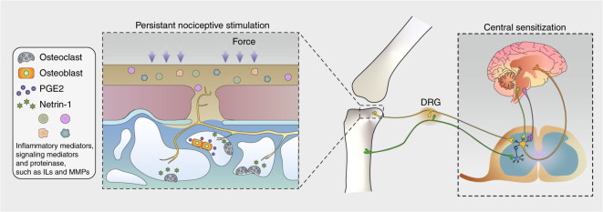
Osteoarthritis comprises several joint disorders characterized by articular cartilage degeneration and persistent pain, causing disability and economic burden. The incidence of osteoarthritis is rapidly increasing worldwide due to aging and obesity trends. Basic and clinical research on osteoarthritis has been carried out for decades, but many questions remain unanswered. The exact role of subchondral bone during the initiation and progression osteoarthritis remains unclear. Accumulating evidence shows that subchondral bone lesions, including bone marrow edema and angiogenesis, develop earlier than cartilage degeneration. Clinical interventions targeting subchondral bone have shown therapeutic potential, while others targeting cartilage have yielded disappointing results. Abnormal subchondral bone remodeling, angiogenesis and sensory nerve innervation contribute directly or indirectly to cartilage destruction and pain. This review is about bone-cartilage crosstalk, the subchondral microenvironment and the critical role of both in osteoarthritis progression. It also provides an update on the pathogenesis of and interventions for osteoarthritis and future research targeting subchondral bone.


Conflict of interest statement

The authors declare no competing interests.

Figures

Fig. 1
Fig. 1
Schematic diagram of subchondral bone responding to aberrant mechanical loading and subsequent cartilage destruction. In the early stage of knee OA, bone resorption is overactivated, especially in the BML area. Biological factors, such as TGF-β, orchestrate MSC recruitment and bone formation, along with the generation of vessels and nerve fibers. Osteopetrosis and bone islets can be observed in advanced stages, which induce cartilage degeneration, at least in part, by abnormal load bearing
Fig. 2
Fig. 2
Subchondral bone microstructural transformation during OA progression. Rod-like and plate-like trabeculae are distributed accurately at a proper ratio to disperse stress. Osteogenesis coupled with angiogenesis lead to an increased BMD and conspicuous microstructural changes at the subchondral level. A decrease in the rod/plate ratio and subchondral bone plate thickening result in excessive mechanical support, which is detrimental to chondrocyte metabolism. Vessel-induced digestion of the cartilage matrix from the bottom up also takes part in the progression of cartilage destruction. a Normal state; b early stage with BML and vascularization; c progressive stage with cartilage damage; d end stage with severe cartilage destruction and vessel erosion
Fig. 3
Fig. 3
TGF-β signaling in the osteochondral area in OA. Shear force breaks the spatial structure of latent TGF-β, thus releasing activated TGF-β and accelerating bone remodeling. On the other hand, overactivated osteoclasts enable TGF-β expression, recruit MSCs and facilitate angiogenesis. In turn, bone and vessel formation promote cartilage TGF-β mobilization, and the cycle continues
Fig. 4
Fig. 4
Coupled angiogenesis and osteogenesis in the subchondral bone accelerate cartilage destruction. Activated TGF-β and PDGF-BB derived from pre-OCs lead to neovascularization, type H vessels couple angiogenesis and osteogenesis by Notch signaling, and OBs accelerate this cycle via RANKL and VEGF release. Endothelial cells digest cartilage and contribute to chondrocyte hypertrophy, which in turn attracts vascular invasion by VEGF signaling
Fig. 5
Fig. 5
Schematic diagram of OA pain. Osteoclast-derived netrin-1 guides sensory nerve innervation in the osteochondral area, and multiple signaling mediators give rise to persistent nociceptive stimulation. The continuous input of pain signals causes central sensitization, suppresses the descending inhibitory system, and brings sympathetic referred pain away from the area of the arthritis lesion

Similar articles

Cited by

References

    1. Hunter DJ, Bierma-Zeinstra S. Osteoarthritis. Lancet. 2019;393:1745–1759. doi: 10.1016/S0140-6736(19)30417-9. - DOI - PubMed
    1. Wilson AJ, Murphy WA, Hardy DC, Totty WG. Transient osteoporosis: transient bone marrow edema? Radiology. 1988;167:757–760. doi: 10.1148/radiology.167.3.3363136. - DOI - PubMed
    1. Zanetti M, Bruder E, Romero J, Hodler J. Bone marrow edema pattern in osteoarthritic knees: correlation between MR imaging and histologic findings. Radiology. 2000;215:835–840. doi: 10.1148/radiology.215.3.r00jn05835. - DOI - PubMed
    1. Roemer FW, et al. MRI-detected subchondral bone marrow signal alterations of the knee joint: terminology, imaging appearance, relevance and radiological differential diagnosis. Osteoarthr. Cartil. 2009;17:1115–1131. doi: 10.1016/j.joca.2009.03.012. - DOI - PubMed
    1. Bowes MA, et al. Osteoarthritic bone marrow lesions almost exclusively colocate with denuded cartilage: a 3D study using data from the osteoarthritis initiative. Ann. Rheum. Dis. 2016;75:1852–1857. doi: 10.1136/annrheumdis-2015-208407. - DOI - PubMed

LinkOut - more resources