

Assessment of clinical radiosensitivity in patients with head-neck squamous cell carcinoma from pre-treatment quantitative ultrasound radiomics
- PMID: 33731738
- PMCID: PMC7969626
- DOI: 10.1038/s41598-021-85221-6
Assessment of clinical radiosensitivity in patients with head-neck squamous cell carcinoma from pre-treatment quantitative ultrasound radiomics
Abstract
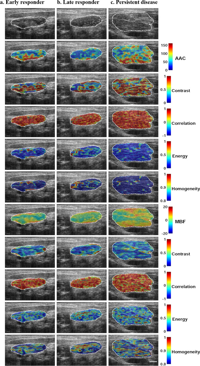
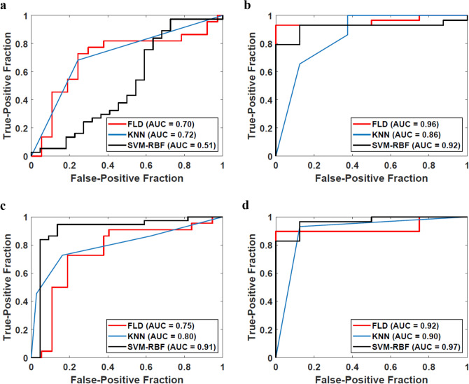
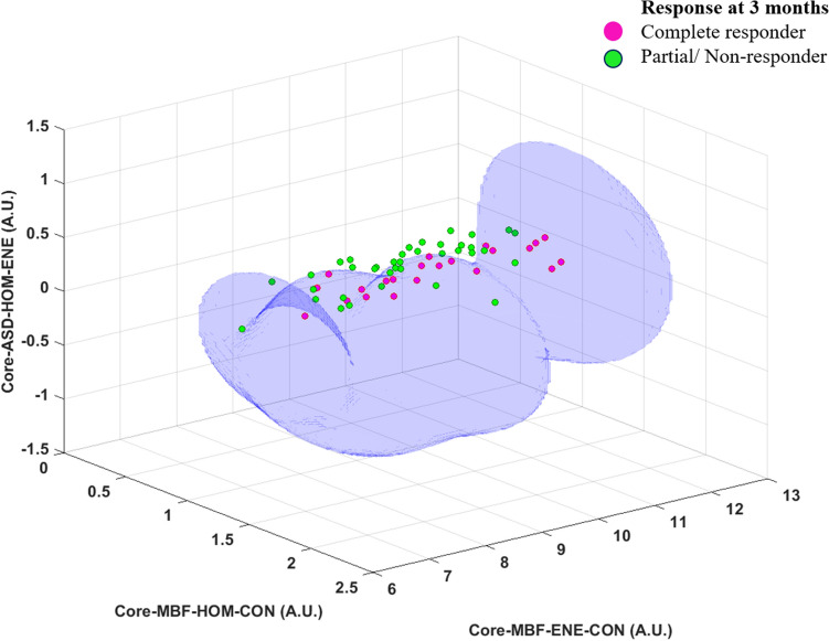
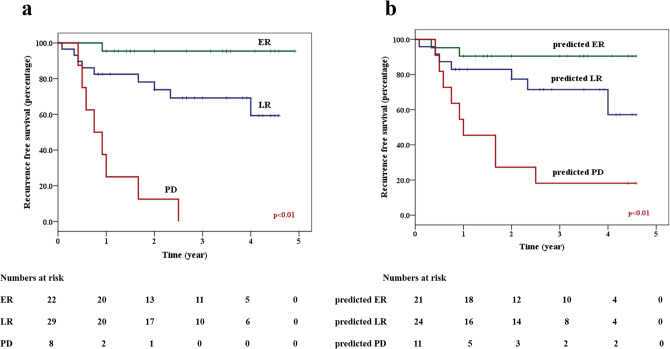
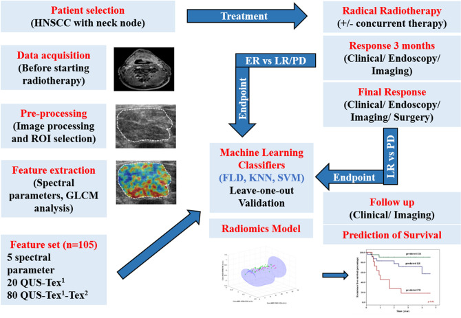
To investigate the role of quantitative ultrasound (QUS) radiomics to predict treatment response in patients with head and neck squamous cell carcinoma (HNSCC) treated with radical radiotherapy (RT). Five spectral parameters, 20 texture, and 80 texture-derivative features were extracted from the index lymph node before treatment. Response was assessed initially at 3 months with complete responders labelled as early responders (ER). Patients with residual disease were followed to classify them as either late responders (LR) or patients with persistent/progressive disease (PD). Machine learning classifiers with leave-one-out cross-validation was used for the development of a binary response-prediction radiomics model. A total of 59 patients were included in the study (22 ER, 29 LR, and 8 PD). A support vector machine (SVM) classifier led to the best performance with accuracy and area under curve (AUC) of 92% and 0.91, responsively to define the response at 3 months (ER vs. LR/PD). The 2-year recurrence-free survival for predicted-ER, LR, PD using an SVM-model was 91%, 78%, and 27%, respectively (p < 0.01). Pretreatment QUS-radiomics using texture derivatives in HNSCC can predict the response to RT with an accuracy of more than 90% with a strong influence on the survival.Clinical trial registration: clinicaltrials.gov.in identifier NCT03908684.
Conflict of interest statement
The authors declare no competing interests.
Figures
Similar articles
-
Quantitative US Delta Radiomics to Predict Radiation Response in Individuals with Head and Neck Squamous Cell Carcinoma.Radiol Imaging Cancer. 2024 Mar;6(2):e230029. doi: 10.1148/rycan.230029. Radiol Imaging Cancer. 2024. PMID: 38391311 Free PMC article.
-
Quantitative ultrasound radiomics in predicting recurrence for patients with node-positive head-neck squamous cell carcinoma treated with radical radiotherapy.Cancer Med. 2021 Apr;10(8):2579-2589. doi: 10.1002/cam4.3634. Epub 2020 Dec 13. Cancer Med. 2021. PMID: 33314716 Free PMC article.
-
Ultrasound delta-radiomics during radiotherapy to predict recurrence in patients with head and neck squamous cell carcinoma.Clin Transl Radiat Oncol. 2021 Mar 12;28:62-70. doi: 10.1016/j.ctro.2021.03.002. eCollection 2021 May. Clin Transl Radiat Oncol. 2021. PMID: 33778174 Free PMC article.
-
Survival outcomes for head and neck cancer patients with N3 cervical nodal metastases.Clin Otolaryngol. 2020 May;45(3):342-349. doi: 10.1111/coa.13501. Epub 2020 Feb 20. Clin Otolaryngol. 2020. PMID: 31869000
-
Chemo-reirradiation in persistent/recurrent head and neck cancers.Jpn J Clin Oncol. 2004 Feb;34(2):61-8. doi: 10.1093/jjco/hyh017. Jpn J Clin Oncol. 2004. PMID: 15067097 Review.
Cited by
-
Application of Machine Learning Methods to Improve the Performance of Ultrasound in Head and Neck Oncology: A Literature Review.Cancers (Basel). 2022 Jan 28;14(3):665. doi: 10.3390/cancers14030665. Cancers (Basel). 2022. PMID: 35158932 Free PMC article. Review.
-
Quantitative US Delta Radiomics to Predict Radiation Response in Individuals with Head and Neck Squamous Cell Carcinoma.Radiol Imaging Cancer. 2024 Mar;6(2):e230029. doi: 10.1148/rycan.230029. Radiol Imaging Cancer. 2024. PMID: 38391311 Free PMC article.
-
The Use of Artificial Intelligence in Head and Neck Cancers: A Multidisciplinary Survey.J Pers Med. 2024 Mar 25;14(4):341. doi: 10.3390/jpm14040341. J Pers Med. 2024. PMID: 38672968 Free PMC article.
-
Radiomics approach for identifying radiation-induced normal tissue toxicity in the lung.Sci Rep. 2024 Oct 16;14(1):24256. doi: 10.1038/s41598-024-75993-y. Sci Rep. 2024. PMID: 39415029 Free PMC article.
-
Early Changes in Quantitative Ultrasound Imaging Parameters during Neoadjuvant Chemotherapy to Predict Recurrence in Patients with Locally Advanced Breast Cancer.Cancers (Basel). 2022 Feb 28;14(5):1247. doi: 10.3390/cancers14051247. Cancers (Basel). 2022. PMID: 35267555 Free PMC article.
References
Publication types
MeSH terms
Associated data
Grants and funding
LinkOut - more resources
Full Text Sources
Other Literature Sources
Medical
