. 2021 Mar 17;11(1):6166.
doi: 10.1038/s41598-021-85360-w.

D-Aspartate consumption selectively promotes intermediate-term spatial memory and the expression of hippocampal NMDA receptor subunits

Affiliations

D-Aspartate consumption selectively promotes intermediate-term spatial memory and the expression of hippocampal NMDA receptor subunits

Gergely Zachar et al. Sci Rep. .

Abstract

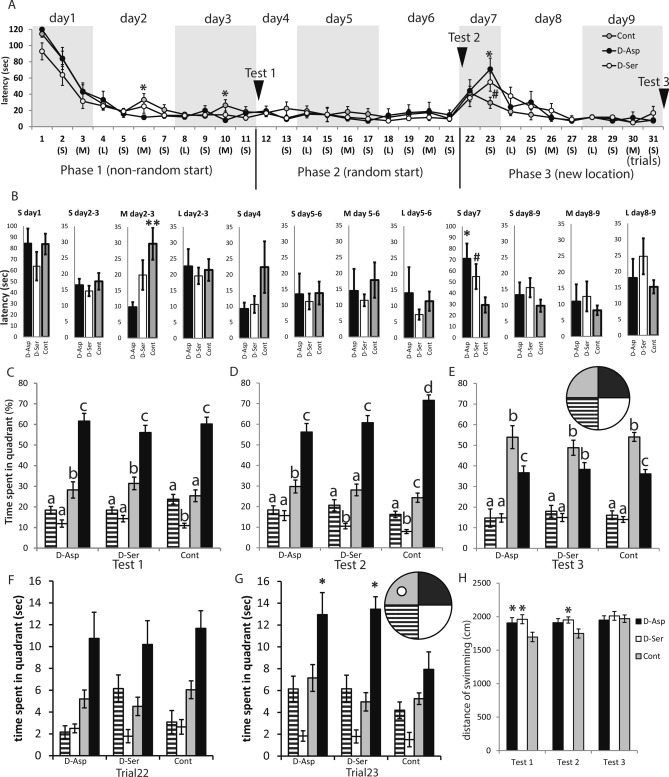
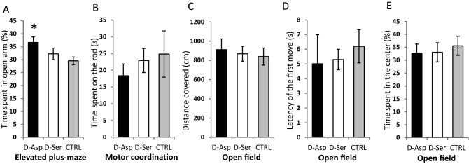
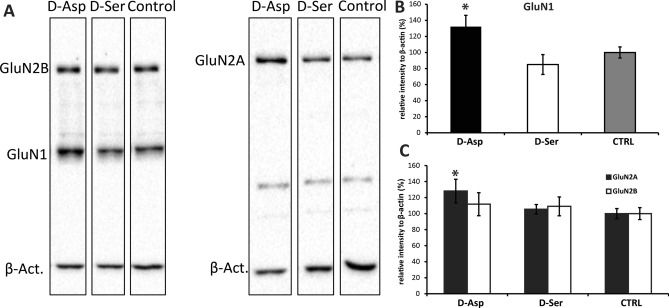
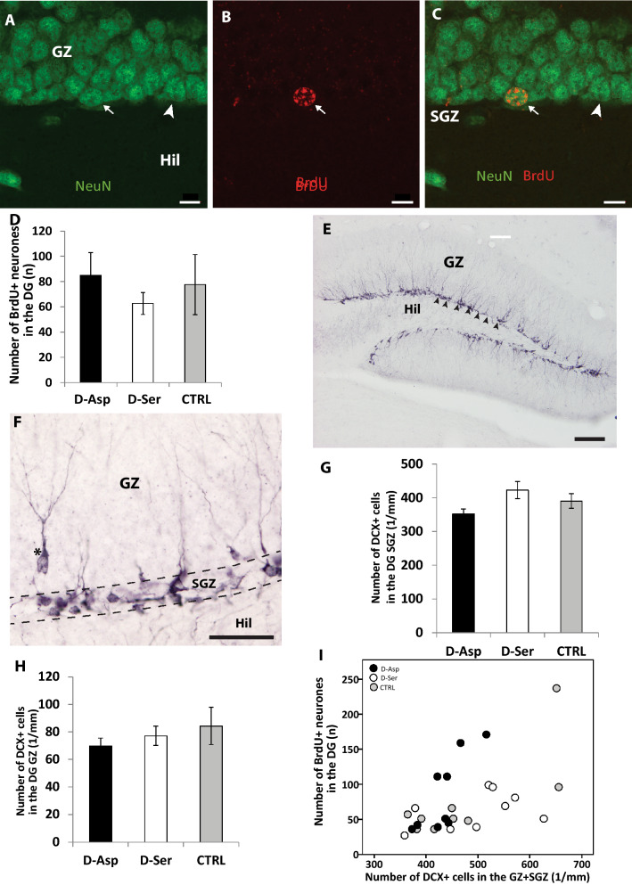
D-Aspartate (D-Asp) and D-serine (D-Ser) have been proposed to promote early-phase LTP in vitro and to enhance spatial memory in vivo. Here, we investigated the behavioural effects of chronic consumption of D-Asp and D-Ser on spatial learning of mice together with the expression of NMDA receptors. We also studied the alterations of neurogenesis by morphometric analysis of bromo-deoxyuridine incorporating and doublecortin expressing cells in the hippocampus. Our results specify a time period (3-4 h post-training), within which the animals exposed to D-Asp (but not D-Ser) show a more stable memory during retrieval. The cognitive improvement is due to elimination of transient bouts of destabilization and reconsolidation of memory, rather than to enhanced acquisition. D-Asp also protracted reversal learning probably due to reduced plasticity. Expression of GluN1 and GluN2A subunits was elevated in the hippocampus of D-Asp (but not D-Ser) treated mice. D-Asp or D-Ser did not alter the proliferation of neuronal progenitor cells in the hippocampus. The observed learning-related changes evoked by D-Asp are unlikely to be due to enhanced proliferation and recruitment of new neurones. Rather, they are likely associated with an upregulation of NMDA receptors, as well as a reorganization of receptor subunit assemblies in existing hippocampal/dentate neurons.


Conflict of interest statement

The authors declare no competing interests.

Figures

Figure 1
Figure 1
Graphical illustration of the experimental timeline of two cohorts of mice: (A) In Experiment 1, spatial learning ability of the mice was assessed using MWM test from day 43 to day 51. Black arrowheads denote the training trials while red arrowheads show the test trials (where the platform was not present). Red arrows represent the time position of trials within a day, along with the preceding intertrial intervals (trial types: M, L or S). EPM elevated plus maze test, *days of the MWM training as shown on Fig. 2. (B) In Experiment 2, BrdU was injected on day 24 of DAA consumption to label proliferating neurones. One day before perfusion open field test followed by a motor coordination test (rod grip test) were carried out on all experimental groups.
Figure 2
Figure 2
(A) Latency of finding the platform in the Morris water-maze test. The training consisted of three distinct phases over the span of 9 days: (1) When the mice were put into the maze consistently at the same point of the basin to start their exploration (non-random start), (2) when their position was chosen randomly around the edge of the maze (random start), and (3) when the platform was relocated to another quadrant (new location). In phase 1, d-Asp treated mice reached the platform faster than control individuals when a medium amount of time (3–4 h) passed after the previous trial (M) but there was no such difference in the latency either in case of a short (S) (10–15 min), or a long (L) (14–16 h) intertrial period. In phase 2, the random starting position did not affect the performance of the individuals in any of the treatment groups. In phase 3, both d-Ser and d-Asp mice were slower than controls to reach the platform when its location was changed. Asterisk (*) represents significant difference between d-Asp and Control (P < 0.05; Tukey post-hoc test). Hash (#) represents significant difference between d-Ser and Control (P = 0.064; Tukey post-hoc test). (B) Diagrams representing the mean latencies of S, M and L trials (as defined in Fig. 1A) in the acquisition period (days 1, 4, 7) or consolidation period (combining days 2–3, 5–6, 8–9). The advantage of the d-Asp individuals in learning the location of the platform was only detectable during phase 1 of learning, in the consolidation period (on days 2–3 of training), in those trials that followed the previous trial after 180–240 min (M). When the platform was relocated to a different quadrant of the water maze, both d-Ser and d-Asp individuals were slower to adapt to the change, however they differed from the control animals only on day 7, at the second trial (10 min after the first trial with the new location). A transient delay of reversal learning with both d-Ser and d-Asp was observed on day 7 (S). *P < 0.05, **P < 0.01, #P = 0.064. (CE) To show that the animals indeed learn the position of the platform (black quadrant) and not using any direct cues to detect it, we tested every animal without the platform after all the three phases of the trainings. The different letters above the columns denote significant difference between the time spent in the quadrants within one treatment group. All mice learned the spatial location of the platform, and spent more time in the appropriate quadrant even with the platform removed. During the reversal (E) d-Asp treated and control animals preferred the new location of the platform (grey quadrant), although the previous location remained their second preferred choice. d-Ser treated mice also tended to prefer the new location over the previous one, however their preference just failed to reach the level of statistical significance (t = 2.47, P = 0.082). (FG) During the second (but not the first, F) trial of the reversal learning (G), both d-Asp and d-Ser individuals spent longer time in the quadrant of previously learned platform location when compared with control mice (d-Asp: t = 2.13, P = 0.039; d-Ser: t = 2.54, P = 0.015). Gray quadrant: new target zone, black quadrant: previous target zone. *P < 0.05 compared to control. (H) Both d-Asp (t = 2.13, P = 0.040) and d-Ser (t = 2.90, P = 0.006) mice covered more distance while swimming than the control animals, in the first platformless test (F2,38 = , P = 0.014). However, during the second test (F2,38 = 3.21, P = 0.05) session, only d-Ser differed from the controls (t = 2.38, P = 0.023). There was no significant difference during the third test session. *P < 0.05 compared to control.
Figure 3
Figure 3
(A) d-Asp treated mice spent more time in the open arm of the elevated plus-maze than control animals (t = 2.61, P = 0.014). *P < 0.05. (B) Neither of the experimental treatment groups differed from the control when tested for motor coordination by a rod grip test (F2,27 = 1.27, P = 0.297). (C) There was no significant difference in the locomotor activity of the treatment groups in an open field test (F2,28 = 1,98, P = 0.157). (D) There was no difference between the groups regarding the latency of the first movement after placing the mouse in the middle of arena (F2,28 = 0.901, P = 0.418). (E) There was no difference between the treatment groups in the time spent at the center of the open-field arena (F2,28 = 0.18, P = 0.838).
Figure 4
Figure 4
(A) Representative blotting membranes showing the position and density of the subunits of NMDARs in the crude synaptosome fraction of hippocampal tissue extracts. The GluN2A and GluN2B subunits were run on different membranes for technical reasons. The bands corresponding to respective GluN subunits and to beta-actin were developed using different exposure times prior to scanning. Region of the blot containing GluN2B subunits has been cut off and processed separately. For full length blots see supplementary Figures S1 and S2. (B) The expression of GluN1 subunit was significantly elevated in the hippocampus of d-Asp consuming mice as compared to controls (*t = 2.05, P = 0.048). (C) d-Asp mice also showed greater expression of the GluN2A subunit as compared to controls (*t = 2.32, P = 0.026), but there was no significant difference in the expression of GluN2B.
Figure 5
Figure 5
(AC) Confocal laser scanning photomicrographs of the granular layer of the dentate gyrus immunolabelled against NeuN (green) and BrdU (red). A-NeuN labelled granule cells visualized using the green channel, (B) A BrdU containing cell nucleus in the same region (red channel), (C) Combined images of A and B to demonstrate that the BrdU + nucleus belongs to a NeuN + neuron. Arrow: BrdU + /NeuN + doubly labelled neurone, Arrowhead: NeuN + neurone, Scale bar: 10 µm. Abbreviations: GZ-granule cell zone, Hil – hilus, SGZ – subgranular zone. (D) Calculated number of newly generated (BrdU +) cells of verified neuronal character (colocalizing with NeuN +) in the given reference volume of dentate gyrus. The treatments had no effect on the number of BrdU + neurones. (F2,24 = 0.445, P = 0.646). (E) Photomicrograph of the dentate gyrus immunolabelled for doublecortin (DCX). The somata and processes of migrating precursor cells are visible at the granular/subgranular border. Arrowheads mark the cells lining up in the subgranular zone, GZ-granule cell zone, Hil-hilus. Scale bar: 200 µm. (F) DCX + cells tends to accumulate at the subgranular proliferative zone (SGZ) of the dentate gyrus, however some of the newly proliferated cells (asterisk) already started to migrate into the granular zone (GZ). Hil-hilus. Scale bar: 50 µm. (GH) Number of DCX + cells measured in a reference volume of the dentate gyrus of mice treated with d-amino acids. The chronic consumption of neither d-Asp (Tukey: P = 0.419) nor d-Ser (Tukey: P = 0.526) facilitated the production of DCX + neurones, compared to controls, in the subgranular (G) (F2,28 = 1.482, P = 0.220) and granular (H) (F2,28 = 0.558, P = 0.578) zone. (I) The number of BrdU + neurones and DCX + cells show positive correlation in the dentate gyrus (R2 = 0.536, n = 27, P = 0.004).

Similar articles

Cited by

References

    1. Errico F, Napolitano F, Nisticò R, Centonze D, Usiello A. d-aspartate: an atypical amino acid with neuromodulatory activity in mammals. Rev. Neurosci. 2009;20:429–440. doi: 10.1515/REVNEURO.2009.20.5-6.429. - DOI - PubMed
    1. McDonald JW, Johnston MV. Physiological and pathophysiological roles of excitatory amino acids during central nervous system development. Brain Res. Brain Res. Rev. 1990;15:41–70. doi: 10.1016/0165-0173(90)90011-C. - DOI - PubMed
    1. Yang H, Zheng G, Peng X, Qiang B, Yuan J. d-Amino acids and d-Tyr-tRNA(Tyr) deacylase: stereospecificity of the translation machine revisited. FEBS Lett. 2003;552:95–98. doi: 10.1016/S0014-5793(03)00858-5. - DOI - PubMed
    1. Fujii N. d-amino acids in living higher organisms. Orig. Life Evol. Biosph. 2002;32:103–127. doi: 10.1023/A:1016031014871. - DOI - PubMed
    1. Errico F, et al. D-aspartate prevents corticostriatal long-term depression and attenuates schizophrenia-like symptoms induced by amphetamine and MK-801. J. Neurosci. 2008;28:10404–10414. doi: 10.1523/JNEUROSCI.1618-08.2008. - DOI - PMC - PubMed

Publication types

Substances

LinkOut - more resources