. 2021 Sep;15(9):2614-2626.
doi: 10.1038/s41396-021-00953-7. Epub 2021 Mar 17.

Nutrient complexity triggers transitions between solitary and colonial growth in bacterial populations

Affiliations

Nutrient complexity triggers transitions between solitary and colonial growth in bacterial populations

Glen G D'Souza et al. ISME J. 2021 Sep.

Abstract

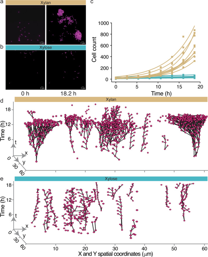
Microbial populations often experience fluctuations in nutrient complexity in their natural environment such as between high molecular weight polysaccharides and simple monosaccharides. However, it is unclear if cells can adopt growth behaviors that allow individuals to optimally respond to differences in nutrient complexity. Here, we directly control nutrient complexity and use quantitative single-cell analysis to study the growth dynamics of individuals within populations of the aquatic bacterium Caulobacter crescentus. We show that cells form clonal microcolonies when growing on the polysaccharide xylan, which is abundant in nature and degraded using extracellular cell-linked enzymes; and disperse to solitary growth modes when the corresponding monosaccharide xylose becomes available or nutrients are exhausted. We find that the cellular density required to achieve maximal growth rates is four-fold higher on xylan than on xylose, indicating that aggregating is advantageous on polysaccharides. When collectives on xylan are transitioned to xylose, cells start dispersing, indicating that colony formation is no longer beneficial and solitary behaviors might serve to reduce intercellular competition. Our study demonstrates that cells can dynamically tune their behaviors when nutrient complexity fluctuates, elucidates the quantitative advantages of distinct growth behaviors for individual cells and indicates why collective growth modes are prevalent in microbial populations.


Conflict of interest statement

The authors declare no competing interests.

Figures

Fig. 1
Fig. 1. The polymer xylan limits the growth of C. crescentus compared to the monomer xylose.
aCaulobacter crescentus CB15 cells were grown in the same concentration (%weight/volume) of the polymer 0.05% xylan or its constituent monomer 0.05% xylose and b the growth dynamics of populations (optical density at 600 nm) were measured. c Maximum growth rate and d maximum optical density observed over the course of a growth cycle. Compared to populations on xylan, populations grown on xylose achieve higher growth rates (h−1) (independent samples t-test, P = 0.0019, R2 (eta2) = 0.82, npopulations = 4 for each treatment) and greater maximum optical density (independent samples t-test, P = 0.0001, R2 (eta2) = 0.94, npopulations = 4 for each treatment). Squares, horizontal lines and whiskers indicate the individual measurements for each biological replicate population (npopulations = 4), the mean and the 95% confidence interval (CI), respectively, on xylan (yellow) and xylose (blue). Asterisks indicate significant differences.
Fig. 2
Fig. 2. Cells display solitary behavior on xylose and aggregative behavior on xylan.
Representative images of C. crescentus CB15 cells (labeled with constitutively expressed mKate2, false colored as magenta) at different time points within the microfluidic growth chambers supplied with either xylan (a) or xylose (b) as the sole source of carbon. c On xylan (yellow), the number of sessile cells in the growth chamber increases with time, whereas on xylose (blue) it remains nearly constant. Squares indicate the number of cells present at a given time point in each chamber (nchambers = 9), with a linear or exponential regression line for each chamber (xylose, linear regression model, R2 = 0.69–0.92, slope = 1.22–3.27, P < 0.01; xylan, exponential growth model, R2 = 0.92–0.99, doubling time = 2.89–4.15 h). Lineage trees reconstructed from time-lapse images of cells within one chamber, for xylan (d) and xylose (e). Cells (magenta spheres) are plotted as a function of their spatial location (x and y axes) and time t. Black lines connect cells that are related through cell division, and branching points mark division events. Representative time-lapse images of cells in xylan and xylose are shown in Supplementary Videos 1 and 2, respectively. Additional lineage trees from the other chambers are depicted in Supplementary Figs. 3 and 4 and a visual representation of lineage development in one representative xylan-fed chamber is shown in Supplementary Video 3.
Fig. 3
Fig. 3. Aggregative behavior results in an increase in cell growth in xylan within microfluidic chambers.
Median single cell growth rates (h−1) of C. crescentus CB15 cells as a function of the number of cells in a chamber on (a) xylan and (b) xylose. After binning cells based on their birth times (bins: 0–2.66 h, 2.67–5.33 h, 5.34–8 h, 8.1–10.66 h, 10.67–13.33 h, 13.34–16 h, 16.1–18.6 h) and hence the number of cells present during their growth, we determined which non-linear regression model can best predict (based on the R2 fit, see Methods and Supplementary Methods for detailed description) the relationship between median growth rate and cell number. Squares represent data for a single bin from one chamber (yellow: xylan, blue: xylose), and lines indicate the trajectory of growth rates for each chamber. In xylan (a), the relationship between median growth rate and the number of cells within a chamber was best explained by an exponential growth model (nchambers = 9, R2 = 0.67–0.83). Maximum growth rates were reached when the number of cells chambers reached 40–110. In xylose (b), median growth rates also increased with the density in the chamber and were also best explained by an exponential growth model (nchambers = 9, R2 = 0.72–84). An analysis of covariance further revealed that there are significant differences in growth rate with cellular density when birth-time is used as a covariate in the xylan environment (F1,6 = 12.37, P < 0.01, R2 = 0.56, eta2 = 0.49) but not the xylose environment (F1,6 = 2.12, P > 0.05, R2 = 0.24, eta2 = 0.18). c In xylan, on average a four-fold lower slope of growth rate with number of cells present in the chamber is observed compared to that in xylose (Mann–Whitney test, P = 0.0006, R2 (eta squared) = 0.76, nchambers = 9 for each treatment). In c, box plots extend from the 25th to 75th percentiles and whiskers indicate the 10th (bottom) and 90th (top) percentiles of median growth rates. Asterisks indicate statistically significant differences between groups, respectively. Also see Supplementary Fig. 7c for a plot of change in growth rate per cell in xylan and xylose. d Initial cell density (cfu ml−1) has a stronger influence on time to reach half maximum optical density in xylan than in xylose, in well-mixed C. crescentus CB 15 populations. This is indicated by a higher slope of linear regression of the times to reach half maximum optical density on xylan compared to xylose (semi-log regression model, xylan: R2 = 0.93, slope = −6.78 h per 10 cells; xylose: R2 = 0.77, slope = −3.41 h per 10 cells; slopes differ significantly, P < 0.05, npopulations = 4). Squares indicate the measurements for each biological replicate (n = 4) and lines show the fit of the regression model. Also see Supplementary Fig. 7f for the relationship between growth rate and inoculum density.
Fig. 4
Fig. 4. The activity of xylanase is localized on individual cells.
ac Xylanase activity (visualized using the degradation of a chromogenic analog of xylan) was present in cells growing on xylan (left panel) and negligible in cells growing on xylose (right panel). Representative (a) phase contrast, (b) fluorescence and (c) merged images of C. crescentus CB15 cells in one chamber that were grown for 18 h on xylose or xylan. Contrasts were adjusted to improve optical clarity but not for measurements in the images. d Mean fluorescence intensities (arbitrary units: a.u.) measured within cells, in their immediate vicinity (the extracellular region closest to the boundary of a cell) and in the background (a region without any cells). Points show the mean intensities for five cells and five corresponding extracellular and background regions each in five different microfluidic chambers, and horizontal lines show the mean and 95% CI. Asterisks and ns denote significant and non-significant differences between groups, respectively (independent samples t-test, FDR corrected q < 0.05, nchambers = 5, ncells/objects = 25).
Fig. 5
Fig. 5. Cells transition between aggregative and solitary behaviors in response to change in nutrient complexity.
ad Time-lapse images of C. crescentus CB15 cells (labeled with constitutively expressed mKate2, false colored as magenta) within chambers exposed to constant conditions or switches in the complexity of the nutrients. e Cell density time series obtained from high frequency (1 frame per min) imaging of one chamber indicates that cell density increases while growing on xylan and starts declining ~40 min after the transition from xylan (Xn) to xylose (Xy). Switching time is indicated by the shaded background. Density is quantified as the number of cells in the area defined by the smallest rectangle encompassing each colony (illustrated by the figure inset), based on the (x,y) coordinates of the cells before the nutrient switch. See Supplementary Video 9 for a time-lapse of cells. f Cell density time series for different nutrient switches (at time 0 h; shaded background) based on images acquired every 8 min, each normalized to the colony density 4 h before the switch. Time series were averaged over nchambers per condition (colored lines) and shown with the corresponding 95% confidence intervals (gray areas). Cells exposed to a xylan-to-xylan mock-switch continued their gradual increase in density (yellow, nchambers = 10), while cells exposed to a xylose-to-xylose mock-switch did not change in average density (blue, nchambers = 9, one chamber excluded because of very low initial density). Switching from xylan to xylose was followed by a decrease in density (red, nchambers = 9), while little change in density was observed when switching from xylose to xylan (black, nchambers = 9). See associated Supplementary Videos 10–12.

References

    1. Nadell CD, Drescher K, Foster KR. Spatial structure, cooperation and competition in biofilms. Nat Rev Microbiol. 2016;14:589–600. doi: 10.1038/nrmicro.2016.84. - DOI - PubMed
    1. Singh PK, Bartalomej S, Hartmann R, Jeckel H, Vidakovic L, Nadell CD, et al. Vibrio cholerae combines individual and collective sensing to trigger biofilm dispersal. Curr Biol. 2017;27:3359–e7. doi: 10.1016/j.cub.2017.09.041. - DOI - PMC - PubMed
    1. D’Souza G, Shitut S, Preussger D, Yousif G, Waschina S, Kost C. Ecology and evolution of metabolic cross-feeding interactions in bacteria. Nat Prod Rep. 2018;35:455–88. doi: 10.1039/C8NP00009C. - DOI - PubMed
    1. McDougald D, Rice SA, Barraud N, Steinberg PD, Kjelleberg S. Should we stay or should we go: mechanisms and ecological consequences for biofilm dispersal. Nat Rev Microbiol. 2012;10:39–50. doi: 10.1038/nrmicro2695. - DOI - PubMed
    1. Leventhal Gabriel E, Martin Ackermann, Schiessl Konstanze T. Why microbes secrete molecules to modify their environment: the case of iron-chelating siderophores. J R Soc Interface. 2019;16:20180674. doi: 10.1098/rsif.2018.0674. - DOI - PMC - PubMed

Publication types