. 2021 May;31(5):517-525.
doi: 10.1038/s41422-021-00487-9. Epub 2021 Mar 17.

Structural basis for bivalent binding and inhibition of SARS-CoV-2 infection by human potent neutralizing antibodies

Affiliations

Structural basis for bivalent binding and inhibition of SARS-CoV-2 infection by human potent neutralizing antibodies

Renhong Yan et al. Cell Res. 2021 May.

Abstract

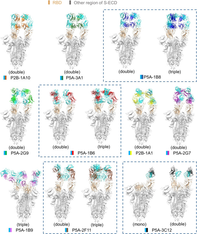
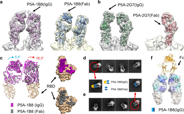
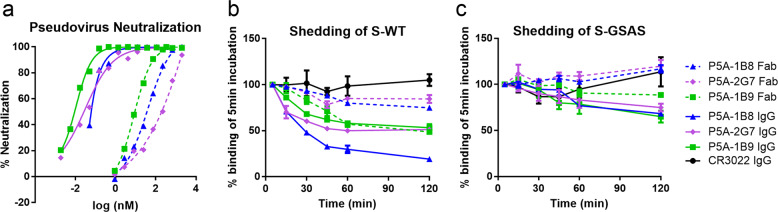
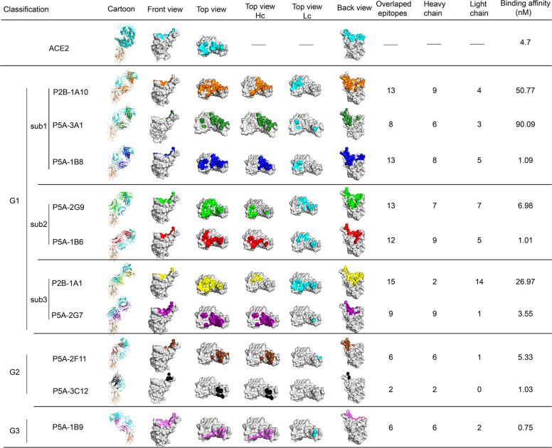
Neutralizing monoclonal antibodies (nAbs) to severe acute respiratory syndrome coronavirus 2 (SARS-CoV-2) represent promising candidates for clinical intervention against coronavirus disease 2019 (COVID-19). We isolated a large number of nAbs from SARS-CoV-2-infected individuals capable of disrupting proper interaction between the receptor binding domain (RBD) of the viral spike (S) protein and the receptor angiotensin converting enzyme 2 (ACE2). However, the structural basis for their potent neutralizing activity remains unclear. Here, we report cryo-EM structures of the ten most potent nAbs in their native full-length IgG-form or in both IgG-form and Fab-form bound to the trimeric S protein of SARS-CoV-2. The bivalent binding of the full-length IgG is found to associate with more RBDs in the "up" conformation than the monovalent binding of Fab, perhaps contributing to the enhanced neutralizing activity of IgG and triggering more shedding of the S1 subunit from the S protein. Comparison of a large number of nAbs identified common and unique structural features associated with their potent neutralizing activities. This work provides a structural basis for further understanding the mechanism of nAbs, especially through revealing the bivalent binding and its correlation with more potent neutralization and the shedding of S1 subunit.


Conflict of interest statement

The authors declare no competing interests.

Figures

Fig. 1
Fig. 1. All solved structures of nAbs in complex with the S protein.
The domain-colored models of all complex are shown. The structures containing different binding modes of the same nAb are boxed with blue dash line. The structures are labeled according to the number of RBD bound with nAb as mono (1 RBD), double (2 RBDs) or triple (3 RBDs) binding, respectively. The heavy chains of IgG are colored in cyan and the light chains of IgG are shown in the indicated colors.
Fig. 2
Fig. 2. Bivalent binding analysis of nAbs.
a Structural comparison between the S/P5A-1B8(IgG) complex and the S/P5A-1B8(Fab) complex. The cryo-EM maps docked with atomic models are shown. b Structural comparison between S/P5A-2G7(IgG) complex and the S/P5A-2G7(Fab) complex. c Comparison between P5A-1B8 (IgG) and P5A-1B8 (Fab) in complex with the S protein. The rotation of the variable region is larger on the right side (18.3°), comparing to the left side(5.4°). Whereas the IgG and the Fab complex have the same epitopes that are colored in purple and gray, respectively. d, e 2D classification of the S/P5A-1B8(IgG) complex and the S/P5A-1B8(Fab) complex re-centered at antibody, respectively. f The cryo-EM map of the S/P5A-1B8(IgG) complex can be docked with the Fc region of a full-length antibody model (PDB code: 5DK3). The Fc, heavy chain, and light chain of the antibody are colored in gold, blue and cyan, respectively.
Fig. 3
Fig. 3. Neutralizing activity and shedding of S1 by IgG- and Fab-forms of nAbs.
a Neutralizing activity against SARS-CoV-2 pseudovirus by P5A-1B8, P5A-1B9, and P5A-2G7 in IgG-forms (solid line) and Fab-forms (dotted line). Data were representative of at least two independent experiments. b Shedding of S1 over time measured using flow cytometry at 37 °C with 293T cell-surface expressed wild-type SARS-Cov-2 S protein. c Similar to b, with a mutant S protein containing GSAS substitution at S1/S2 cleavage site. Data were from three independent experiments, shown as means ± SEM.
Fig. 4
Fig. 4. The classification and epitope of the 10 anti-SARS-CoV-2 nAbs.
The 10 antibodies could be classified into three groups (G1, group 1; G2, group 2; G3, group 3). Group 1 antibodies can be further divided into three subgroups (sub1, sub2, sub3). The complexes of RBD with ACE2 or nAbs are shown as cartoon with RBD colored in wheat, the light chains of nAbs colored in cyan and the heavy chains of nAbs colored in different colors. For the epitope display, RBD is shown as gray surface in top, front and back views, with interface that binds to ACE2 colored in cyan and the epitopes of different nAbs shown in respective colors. For the top views, the epitopes corresponding to heavy chains are shown in respective colors (Top view Hc) and epitopes corresponding to light chains are shown in cyan (Top view Lc). Hc, heavy chain; Lc, light chain. The “Overlapped epitopes” column displays the residue number of nAb epitope that overlaps with ACE2-binding site. The “Heavy chain” and “Light chain” columns show the residue number of the epitope of the respective chain that overlaps with ACE2-binding site. The binding affinity to RBD of IgG antibodies was also shown.

Similar articles

Cited by

References

    1. Wu F, et al. A new coronavirus associated with human respiratory disease in China. Nature. 2020;579:265–269. - PMC - PubMed
    1. Zhu N, et al. A Novel Coronavirus from Patients with Pneumonia in China, 2019. N. Engl. J. Med. 2020;382:727–733. - PMC - PubMed
    1. Ksiazek TG, et al. A novel coronavirus associated with severe acute respiratory syndrome. N. Engl. J. Med. 2003;348:1953–1966. - PubMed
    1. Zaki AM, van Boheemen S, Bestebroer TM, Osterhaus AD, Fouchier RA. Isolation of a novel coronavirus from a man with pneumonia in Saudi Arabia. N. Engl. J. Med. 2012;367:1814–1820. - PubMed
    1. Lan J, et al. Structure of the SARS-CoV-2 spike receptor-binding domain bound to the ACE2 receptor. Nature. 2020;581:215–220. - PubMed

Publication types

MeSH terms