. 2021 Mar;7(3):365-375.
doi: 10.1038/s41477-021-00876-3. Epub 2021 Mar 15.

How the O2-dependent Mg-protoporphyrin monomethyl ester cyclase forms the fifth ring of chlorophylls

Affiliations

How the O2-dependent Mg-protoporphyrin monomethyl ester cyclase forms the fifth ring of chlorophylls

Guangyu E Chen et al. Nat Plants. 2021 Mar.

Abstract

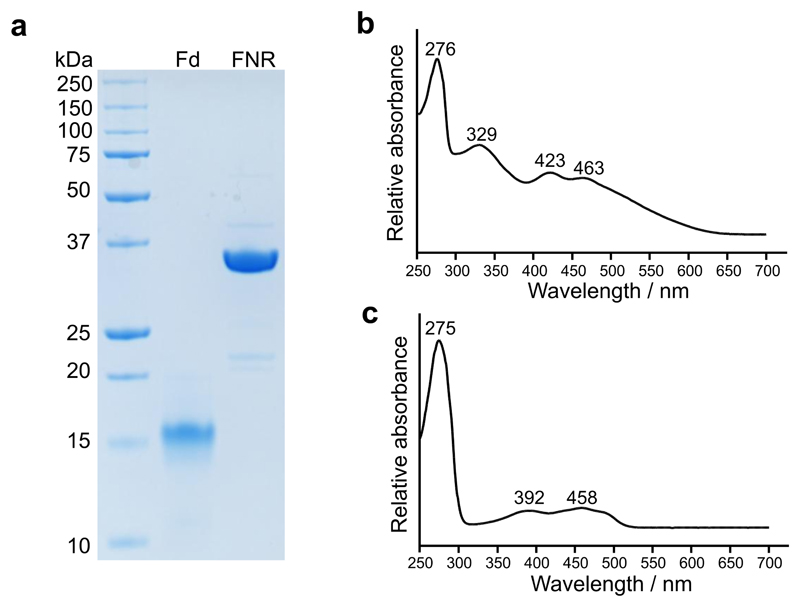
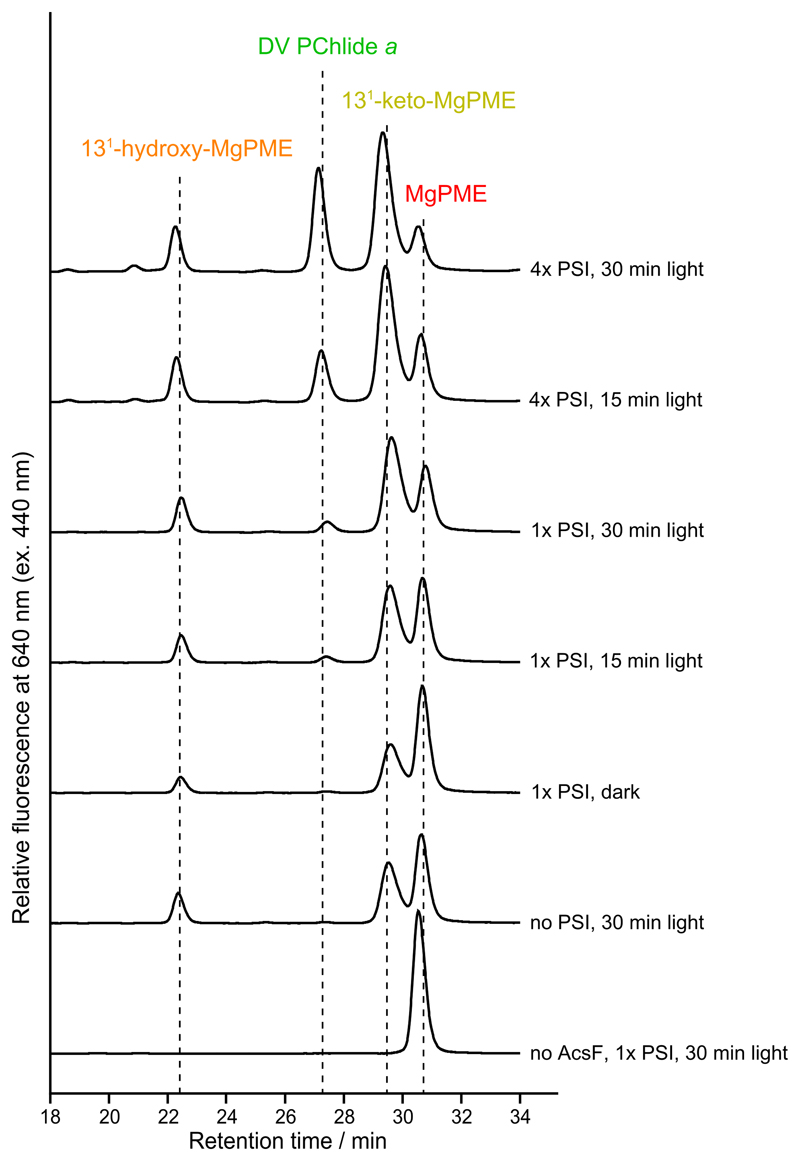
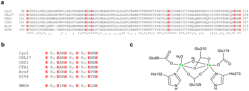
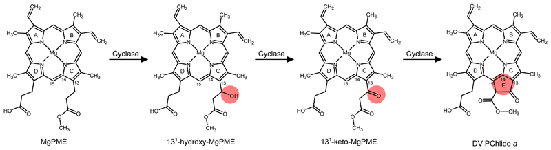
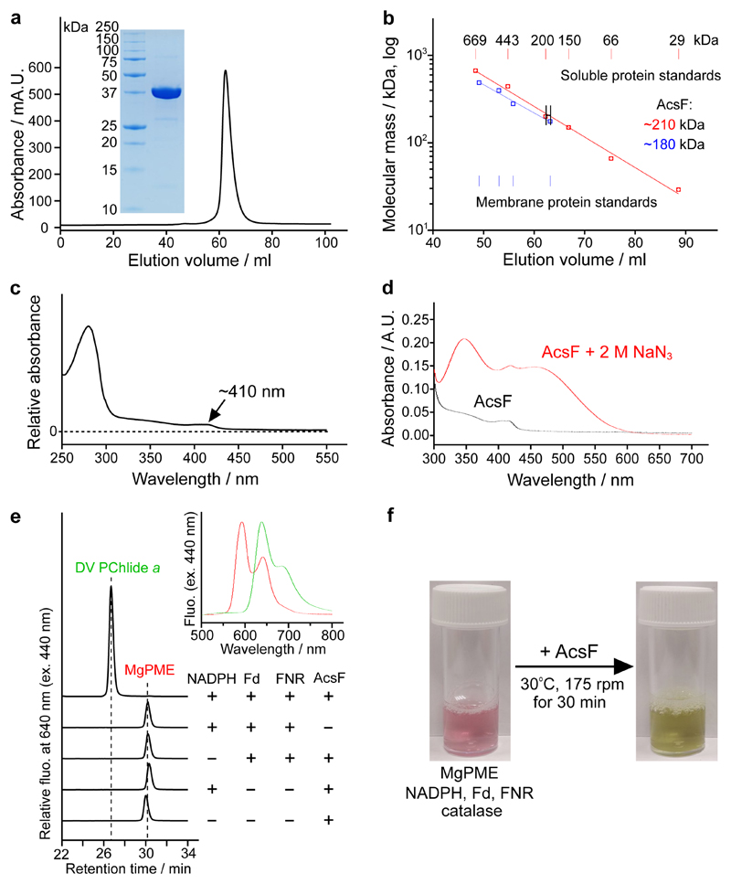
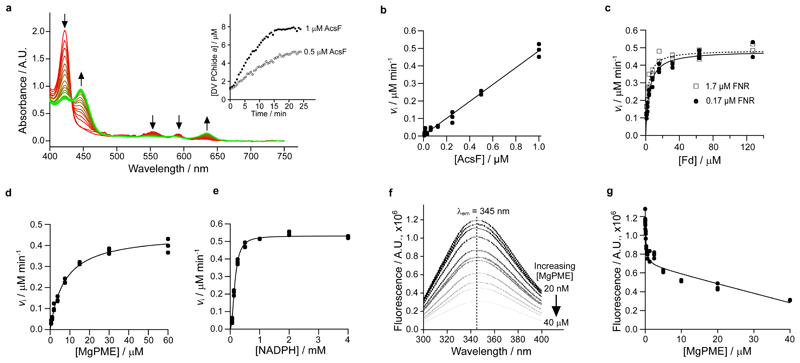
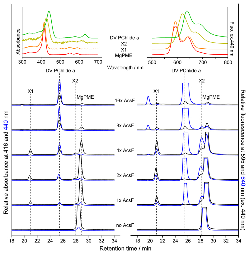
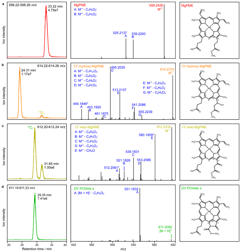
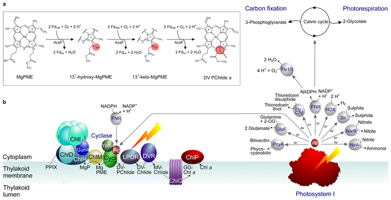
Mg-protoporphyrin IX monomethyl ester (MgPME) cyclase catalyses the formation of the isocyclic ring, producing protochlorophyllide a and contributing substantially to the absorption properties of chlorophylls and bacteriochlorophylls. The O2-dependent cyclase is found in both oxygenic phototrophs and some purple bacteria. We overproduced the simplest form of the cyclase, AcsF, from Rubrivivax gelatinosus, in Escherichia coli. In biochemical assays the di-iron cluster within AcsF is reduced by ferredoxin furnished by NADPH and ferredoxin:NADP+ reductase, or by direct coupling to Photosystem I photochemistry, linking cyclase to the photosynthetic electron transport chain. Kinetic analyses yielded a turnover number of 0.9 min-1, a Michaelis-Menten constant of 7.0 µM for MgPME and a dissociation constant for MgPME of 0.16 µM. Mass spectrometry identified 131-hydroxy-MgPME and 131-keto-MgPME as cyclase reaction intermediates, revealing the steps that form the isocyclic ring and completing the work originated by Sam Granick in 1950.


Conflict of interest statement

Competing interests

The authors declare no competing interests.

Figures

Extended Data Fig. 1
Extended Data Fig. 1. Analysis of purified Anabaena Fd and FNR.
a, SDS-PAGE analysis of purified Anabaena Fd and FNR. b, Absorbance spectrum of purified Anabaena Fd. c, Absorbance spectrum of purified Anabaena FNR.
Extended Data Fig. 2
Extended Data Fig. 2. HPLC elution profiles of pigment extracts from coupled PSI-cyclase assays.
A complete assay contained 2 µM AcsF, 0.04 mg mL-1 spinach Fd, 14 µM MgPME, spinach PSI containing 6 (1x PSI) or 22.4 µM (~4x PSI) Chl a, 20 µM spinach Pc, 2 mM Asc, 60 µM DCPIP and 0.29 mg mL-1 catalase. Assays were incubated either in the dark for 30 min, or under red light illumination for 15 or 30 min. Pigment extracts from the assays were analysed by HPLC and pigment elution was monitored by fluorescence at 640 nm excited at 440 nm. Pigment species were identified by retention times and fluorescence spectra (as in Fig. 4). See Supplementary Fig. 4a for HPLC analysis of pigment extracts from additional control assays.
Extended Data Fig. 3
Extended Data Fig. 3. The diiron binding motif and proposed diiron ligation of AcsF.
a, Sequence alignments showing the conserved diiron binding motif of AcsF proteins. Sequences are from Synechocystis sp. PCC 6803 (CycI, BAA16583), Arabidopsis thaliana (CHL27, NP_191253), Chlamydomonas reinhardtii (CRD1, XP_001692557; CTH1, XP_001691047), Rubrivivax gelatinosus IL144 (AcsF, BAL96694) and Rhodobacter sphaeroides 2.4.1 (0294, abbreviated for RSP_0294, YP_353369). Conserved, highly similar and similar residues are marked with asterisks, colons and full stops, respectively. The putative diiron ligands are in red and bold. Full-length protein sequences were used for alignments but for clarity, only the putative diiron binding motifs with the residue range indicated, are shown. b, Sequence homologies between the diiron binding motifs of AcsF proteins and the soluble methane monooxygenase hydroxylase subunit from Methylococcus capsulatus Bath (MMOH, P22869). c, Proposed coordination of the diiron ligands of AcsF at the diferrous state based on the crystal structure of MMOH (PDB, 1FYZ)51.
Fig. 1
Fig. 1. Proposed reaction intermediates of MgPME cyclase.
Formation of the E ring of chlorophyll is proposed to proceed via hydroxylation, oxidation and cyclisation of the C13 methylpropionyl side chain of MgPME. The chemical change at each step is highlighted. International Union of Pure and Applied Chemistry numbering of the relevant macrocycle carbons is indicated.
Fig. 2
Fig. 2. Purification, spectral characterisation and reconstitution of cyclase activity of AcsF.
a, Gel filtration profile of AcsF on a HiLoad 16/600 Superdex 200 prep grade column monitored by absorbance at 280 nm and SDS-PAGE analysis of 10 µg purified AcsF (inset). Shown are representative of three independently repeated experiments. b, Estimate of the molecular mass of native AcsF from triplicate gel filtration runs (range of elution volume indicated) using calibration curves (logarithm of molecular mass vs elution volume) generated from the data points of soluble (red circles) and membrane (blue squares) protein standards using nonlinear regression analysis. Membrane protein standards of 104, 208, 325 and 416 kDa were used and a value of 72 kDa (size of β-DDM micelles) was added to each molecular mass when generating the calibration curve. The calculated molecular mass values (inset) include the contribution of bound β-DDM molecules. c, Absorbance spectrum of AcsF as isolated. d, Absorbance spectra of 8 µM AcsF in the absence and presence of 2 M sodium azide. e, Cofactor requirements for in vitro cyclase activity of AcsF revealed by end-point HPLC-based assays. A complete assay contained 3.7 µM AcsF, 10 µM MgPME, 2 mM NADPH, 0.2 mg mL-1 spinach Fd, 0.4 units mL-1 spinach FNR and 0.29 mg mL-1 catalase. Retention times and fluorescence spectra (inset) were used to identify pigment species. See Materials and Methods for experimental details. f, Photographs showing the marked colour change showing the activity of AcsF in an assay containing 27 µM MgPME and other assay components at the same concentration as in a.
Fig. 3
Fig. 3. Steady-state kinetics of AcsF, and binding of MgPME to AcsF analysed by tryptophan fluorescence quenching.
a, The progressive spectral change during a continuous absorbance-based cyclase assay, which contained 1 µM AcsF, 10 µM MgPME, 7.6 µM Anabaena Fd, 0.17 µM Anabaena FNR, 2.5 mM NADPH and 0.29 mg mL-1 catalase. Arrows indicate the direction of change. Inset shows the product (DV PChlide a) evolution with 0.5 and 1 µM AcsF, monitored by absorbance at 634 nm. be, The dependence of the initial rate of product formation on AcsF (b), Anabaena Fd (c), MgPME (d) and NADPH (e). Assay conditions were as in a except the following stated differences: b, 7.81 nM to 1 µM AcsF; c, 0.5 µM AcsF, 0.17 or 1.7 µM Anabaena FNR, 0.99 to 127 µM Anabaena Fd; d, 0.5 µM AcsF, 1.7 µM Anabaena FNR, 31 µM Anabaena Fd; e, 0.5 µM AcsF, 30 µM MgPME, 1.7 µM Anabaena FNR, 31 µM Anabaena Fd, 62.5 µM to 4 mM NADPH. Each data point is an independent experiment. The Michaelis-Menten equation (equation 1, see Materials and Methods) was fitted to the kinetic data in c and d, with the characterising parameters K M (apparent) = 4.05 ± 0.39 µM (0.17 µM FNR) or 2.41 ± 0.26 µM (1.7 µM FNR) (c); = 0.91 ± 0.02 min-1, = 7.03 ± 0.51 µM (d). The Hill equation (equation 2, see Materials and Methods) was fitted to the NADPH titration data with = 1.06 ± 0.01 min-1, = 0.16 ± 0.01 mM, n = 2.1 ± 0.1 (e). f, A series of spectra showing quenching of AcsF fluorescence by MgPME. Excitation was set at 280 nm, producing an emission maximum at 345 nm. The average fluorescence spectra of triplicate experiments are shown. g, Plot of AcsF fluorescence against MgPME concentration. Each data point is an independent experiment. The curve fit is described by a modified single-site binding model (equation 3, see Materials and Methods) with K d for MgPME binding of 0.16 ± 0.05 µM.
Fig. 4
Fig. 4. HPLC elution profiles of pigment extracts from end-point cyclase assays at various AcsF concentrations.
Assay conditions were 20 µM MgPME, 0.2 mg mL-1 spinach Fd, 0.4 units mL-1 spinach FNR, 2 mM NADPH, 0.29 mg mL-1 catalase and AcsF at various concentrations from 0.23 (1x AcsF) to 3.68 (16x AcsF) µM. Assays were initiated by adding AcsF and terminated after 30 min incubation. Pigment extracts from the assays were analysed by HPLC with elution of pigment species monitored by absorbance at 416 and 440 nm and by fluorescence at 595 and 640 nm with excitation at 440 nm. Insets show the acquired absorbance and fluorescence spectra of MgPME, DV PChlide a, and the potential reaction intermediates, X1 and X2.
Fig. 5
Fig. 5. Analysis of extracted pigments by LC-ESI-MS/MS.
The pigment extract from scaled-up in vitro cyclase assays corresponding to 4x AcsF (0.92 µM) in Fig. 4 was analysed. Extracted ion chromatograms (EICs) and product ion spectra derived from HCD of selected monoisotopic molecular ions are shown in the left and centre panels, respectively: a, MgPME, b, 131-hydroxy-MgPME, c, 131-keto-MgPME, d, DV PChlide a. The molecular structures that align with the mass spectral evidence presented here are shown in the corresponding right panels. EICs were generated for the indicated m/z ranges covering the target monoisotopic ions with peaks labelled with their retention times and ion intensities. Peaks mapping to 13C-containing isotopomers that fall within the EIC range are also labelled. Cations generated by gas phase neutral loss reactions are indicated by upper case letters with the eliminated molecular formulas also listed. The majority of product ions are carbocations formed after radical neutral loss; those labelled with an asterisk are radical cations formed after even electron neutral loss. Details of the structures which validate the identifications of the cyclase substrate, intermediates and product are shown in Supplementary Fig. 3.
Fig. 6
Fig. 6. Diagram depicting the Fd-dependent cyclase reaction catalysed by AcsF and the supply of reduced Fd, directly or indirectly, by PSI.
a, The updated sequence of cyclase reactions catalysed by AcsF, with the chemical change of the porphyrin substrate at each step highlighted in the pink circle. Fdred and Fdox represent reduced and oxidised Fd, respectively. b, The chlorophyll biosynthesis pathway is shown on the left, progressing from protoporphyrin IX (PPIX) to chlorophyll a (Chl a), via magnesium protoporphyrin IX (MgP), Mg-protoporphyrin IX monomethyl ester (MgPME), 3,8-divinyl protochlorophyllide a (DV-PChlide), divinyl chlorophyllide a (DV-Chlide), monovinyl chlorophyllide a (MV-Chlide), and geranylgeranyl-chlorophyll a (GG-Chl a). ChlH, D, I are subunits, and Gun4 is an accessory protein, of the magnesium chelatase complex; ChlM is the MgP methyltransferase; CycI is the counterpart of the AcsF cyclase in cyanobacteria and plants, shown here with the accessory protein Ycf54 and Fd; LPOR is the light-dependent PChlide oxidoreductase; DVR is the divinyl reductase, the cyanobacterial version (BciB) of which requires Fd, whereas the plant DVR does not. ChlG is the chlorophyll synthase and ChlP is the geranylgeranyl reductase. The diagram (right) depicts a possible direct link between PSI and chlorophyll biosynthesis, showing that PSI could provide reduced Fd for the cyclase reaction; FNR-based reduction of Fd is also depicted, corresponding to the in vitro assays in Fig. 3 and Extended Data Fig. 2 respectively. Reduced Fd also provides electrons for a variety of cellular functions, shown here for cyanobacterial metabolism and adapted from the diagram in ref. . PcyA, phycocyanobilin:Fd oxidoreductase; GlsF, Fd-dependent glutamate synthase; FtrC/V, Fd:thioredoxin reductase; FNR, Fd:NADP+ reductase; Sir, Fd:sulfite reductase; NarB, nitrate reductase; NirA, nitrite reductase; Flv1/3, Flavodiiron 1/3; HOX, bi-directional hydrogenase.

Similar articles

Cited by

References

    1. Porra RJ, Schäfer W, Gad’on N, Katheder I, Drews G, Scheer H. Origin of the two carbonyl oxygens of bacteriochlorophyll a Demonstration of two different pathways for the formation of ring E in Rhodobacter sphaeroides and Roseobacter denitrificans and a common hydratase mechanism for 3-acetyl group formation. Eur J Biochem. 1996;239:85–92. - PubMed
    1. Porra RJ, Urzinger M, Winkler J, Bubenzer C, Scheer H. Biosynthesis of the 3-acetyl and 131-oxo groups of bacteriochlorophyll a in the facultative aerobic bacterium, Rhodovulum sulfidophilum: The presence of both oxygenase and hydratase pathways for isocyclic ring formation. Eur J Biochem. 1998;257:185–191. - PubMed
    1. Wiesselmann M, Hebecker S, Borrero-de Acuña JM, Nimtz M, Bollivar D, Jänsch L, Moser J, Jahn D. Mg-protoporphyrin IX monomethyl ester cyclase from Rhodobacter capsulatus: Radical SAM-dependent synthesis of the isocyclic ring of bacteriochlorophylls. Biochem J. 2020 BCJ20200761. - PubMed
    1. Chen GE, Canniffe DP, Hunter CN. Three classes of oxygen-dependent cyclase involved in chlorophyll and bacteriochlorophyll biosynthesis. Proc Natl Acad Sci USA. 2017;114:6280–6285. - PMC - PubMed
    1. Pinta V, Picaud M, Reiss-Husson F, Astier C. Rubrivivax gelatinosus acsF (previously orf358) codes for a conserved, putative binuclear-iron-cluster-containing protein involved in aerobic oxidative cyclization of Mg-protoporphyrin IX monomethylester. J Bacteriol. 2002;184:746–753. - PMC - PubMed

MeSH terms

Supplementary concepts