. 2021 Apr;592(7854):457-462.
doi: 10.1038/s41586-021-03351-3. Epub 2021 Mar 17.

Expansible residence decentralizes immune homeostasis

Affiliations

Expansible residence decentralizes immune homeostasis

Sathi Wijeyesinghe et al. Nature. 2021 Apr.

Abstract

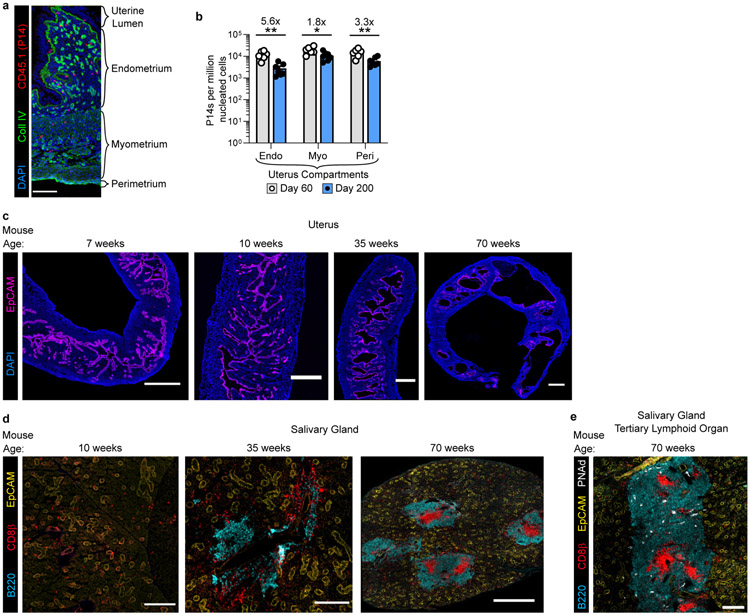
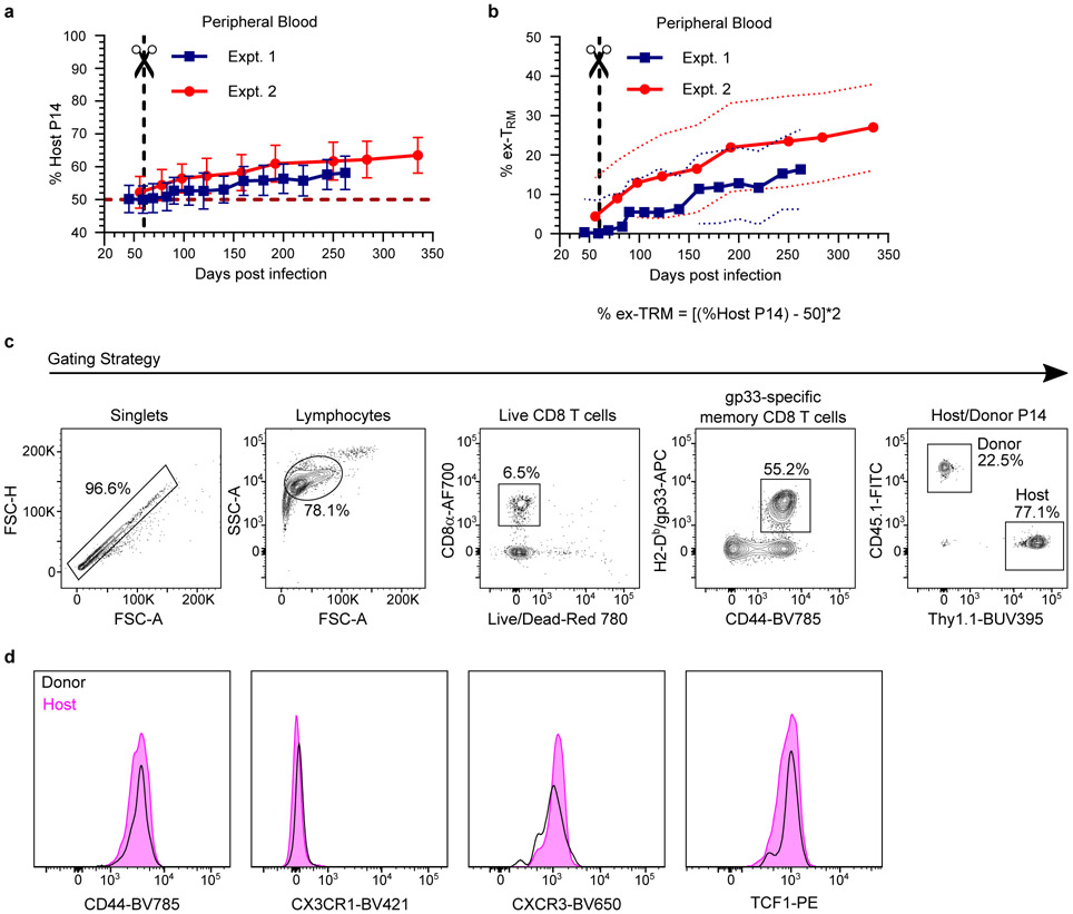
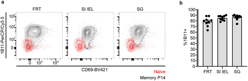
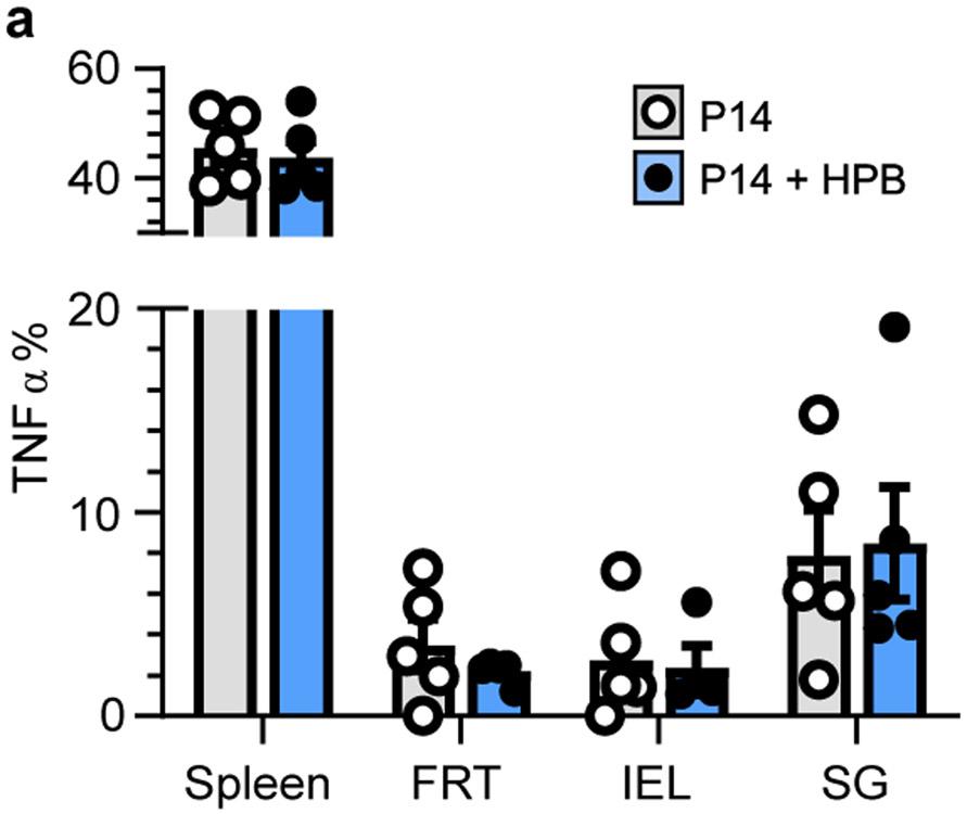
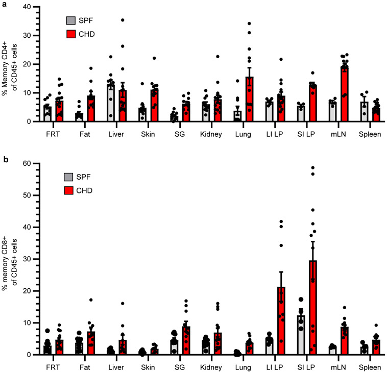
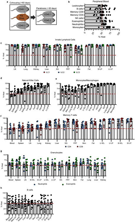
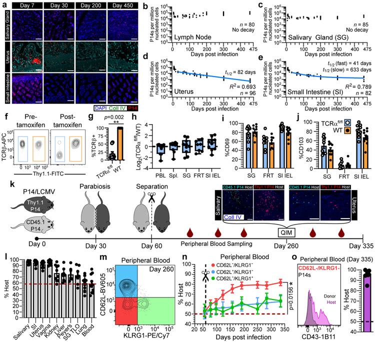
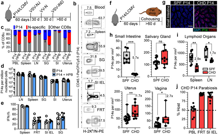
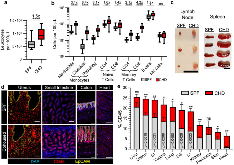
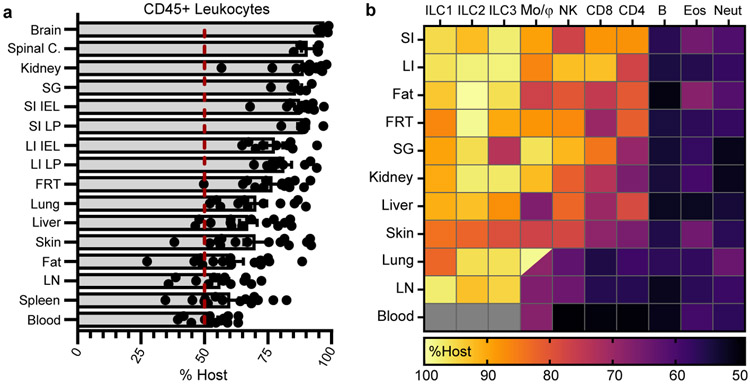
In metazoans, specific tasks are relegated to dedicated organs that are established early in development, occupy discrete locations and typically remain fixed in size. The adult immune system arises from a centralized haematopoietic niche that maintains self-renewing potential1,2, and-upon maturation-becomes distributed throughout the body to monitor environmental perturbations, regulate tissue homeostasis and mediate organism-wide defence. Here we examine how immunity is integrated within adult mouse tissues, and address issues of durability, expansibility and contributions to organ cellularity. Focusing on antiviral T cell immunity, we observed durable maintenance of resident memory T cells up to 450 days after infection. Once established, resident T cells did not require the T cell receptor for survival or retention of a poised, effector-like state. Although resident memory indefinitely dominated most mucosal organs, surgical separation of parabiotic mice revealed a tissue-resident provenance for blood-borne effector memory T cells, and circulating memory slowly made substantial contributions to tissue immunity in some organs. After serial immunizations or cohousing with pet-shop mice, we found that in most tissues, tissue pliancy (the capacity of tissues to vary their proportion of immune cells) enables the accretion of tissue-resident memory, without axiomatic erosion of pre-existing antiviral T cell immunity. Extending these findings, we demonstrate that tissue residence and organ pliancy are generalizable aspects that underlie homeostasis of innate and adaptive immunity. The immune system grows commensurate with microbial experience, reaching up to 25% of visceral organ cellularity. Regardless of the location, many populations of white blood cells adopted a tissue-residency program within nonlymphoid organs. Thus, residence-rather than renewal or recirculation-typifies nonlymphoid immune surveillance, and organs serve as pliant storage reservoirs that can accommodate continuous expansion of the cellular immune system throughout life. Although haematopoiesis restores some elements of the immune system, nonlymphoid organs sustain an accrual of durable tissue-autonomous cellular immunity that results in progressive decentralization of organismal immune homeostasis.


Figures

Extended Data Fig. 1.
Extended Data Fig. 1.. Compartmentalized decay of uterine T cells concomitant with morphological changes in tissue architecture over time.
a, Representative immunofluorescence of uterine tissue. b, The frequency of P14 memory CD8+ T cells in uterine compartments was assessed by quantitative immunofluorescent microscopy at day 60 (n = 6 mice) and day 200 (n = 7 mice) after LCMV infection in one experiment. c, Representative immunofluorescence images of mouse uterine tissue at various ages demonstrating endometrial vacuolations in older mice . d, e, Representative immunofluorescence images of mouse salivary gland at various timepoints demonstrating emergence of salivary gland tertiary lymphoid organs in older mice (d) and expression of peripheral node addressin (PNAd) (e). Morphology representative of n > 12 mice, PNAd staining representative of n = 5 mice (c-e). Coll IV, collagen type IV. 100 μm scale bar (a), 500 μm scale bars (c), 200 μm scale bars (10/35 weeks) and 500 μm scale bar (70 weeks) (d), 200 μM scale bar (e). Statistical significance was determined by two-tailed Mann-Whitney U test (b) where *p = 0.0221, **p = 0.0023 (endometrium) or **p = 0.0082 (perimetrium). Data are presented as mean values +/− SEM.
Extended Data Fig. 2.
Extended Data Fig. 2.. Selective TCR ablation using TCRαfl/fl mice reveals TCR-independent homeostasis of TRM.
a, Experimental model. Thy1.1/CD45.2+ UBC-CreERT2 TCRαfl/fl mice (TCRαfl/fl mice, for brevity) and Thy1.1+/CD45.2+ WT B6 mice were infected with LCMV. After 30 days, 107 lymphocytes, isolated from secondary lymphoid organs, were transferred into naive CD45.1+ B6 mice which were subsequently infected with LCMV. 40 days after infection, CD45.1+ mice were treated with tamoxifen to selectively ablate TCR from transferred Thy1.1/CD45.2+ TCRαfl/fl secondary memory T cells. b, c, LCMV-specific secondary memory T cells in peripheral blood are shown 40 days after LCMV infection (prior to tamoxifen treatment). Data pooled from three independent experiments for a total of n = 8 mice (c). d, e, Selective TCR ablation of TCRαfl/fl secondary memory CD8+ T cells as measured by ex vivo peptide stimulation assay. 60 days after tamoxifen treatment of CD45.1+ B6 recipients, splenocytes were isolated and stimulated in vitro with gp33-41 peptide. Cytokine production by TCR TCRαfl/fl memory CD8+ T cells and TCR+ WT memory CD8+ T cells from spleen is shown and reflects n = 6 mice (d, e). f, The frequency of cells that lack TCRβ expression on TCRαfl/fl memory CD8+ T cells is shown. Data pooled from four independent experiments for a total of n = 8-10 mice (n varies by tissue) (f). g, Representative flow cytometry depicts expression of tissue-resident markers on small intestine epithelial (SI IEL) memory CD8+ T cells 60 days after tamoxifen treatment. h, The frequency of CD69+ memory CD8+ T cells in the spleen is shown for WT and TCRβ TCRαfl/fl populations. Data pooled from four independent experiments for a total of n = 8-10 mice (n varies by tissue) (h). Statistical significance was determined by two-tailed Wilcoxon matched-pairs signed rank test (e, h), where *p = 0.0313. Data are presented as mean values +/− SEM.
Extended Data Fig. 3.
Extended Data Fig. 3.. In vitro activation of TCRαfl/fl naïve T cells generates primary TRM that are maintained in the absence of constitutive TCR signaling.
a, Experimental model. Lymphocytes were isolated from secondary lymphoid organs of CD45.2+ UBC-CreERT2 TCRαfl/fl mice (TCRαfl/fl mice, for brevity) and WT Thy1.1+ B6 mice and enriched for naive CD8+ T cells via magnetic bead enrichment. T cells were activated in vitro for 3 days with anti-CD3ε and rB7-1 and 107 cells were co-transferred into naïve CD45.1+ B6 mice. 30 days later, recipients were treated with tamoxifen. 30 days after tamoxifen treatment, transferred CD8+ T cells were evaluated for CD44 expression, as compared to endogenous CD8+ T cells, shown via representative flow cytometry of CD8+ T cells isolated from blood (b). Expression of TCRβ was evaluated for TCRαfl/fl and WT CD8+ T cells, as shown via representative flow cytometry of peripheral blood (c). The ratio of TCRαfl/fl/WT CD8+ T cells was quantified 30 days after tamoxifen in various tissues, normalized to values from blood, and not significantly different from 1:1. (d). Data show n = 4 biologically independent mice from one experiment. Statistical significance was determined by two-tailed one sample Wilcoxon test using 0 as a hypothetical mean. Data are presented as box plots showing median, IQR, and extrema.
Extended Data Fig. 4.
Extended Data Fig. 4.. CD69 does not unequivocally distinguish long-lived resident memory T cells in the lung.
a-b, Representative flow cytometry (a) and graph (b) demonstrating degree of disequilibrium among CD69+ extravascular memory P14 CD8+ T cells in tissues of separated parabiotic mice (n = 10), 260 days after LCMV infection from one experiment. Upper panels are gated on extravascular memory CD8+ P14 T cells (a). FRT, female reproductive tract. SI, small intestine. IEL, intestinal epithelial lymphocytes. LP, lamina propria. SG, salivary gland. Data are presented as mean values +/− SEM.
Extended Data Fig. 5.
Extended Data Fig. 5.. Ex-TRM comprise a substantial fraction of bloodborne memory.
a-b, Longitudinal graphs depicting the frequency of host-derived memory P14 CD8+ T cells (a) or the frequency of ex-TRM, as calculated (b) in peripheral blood of separated parabiotic mice from two independent experiments (n = 17). Data are presented as mean values +/− SEM and in b, colored dotted lines reflect SEM (b). c-d, >200 days after separation of congenically distinct parabiotic P14 chimeras (n = 17), host- and donor-derived P14 CD8+ T cells were evaluated for expression of markers of antigen experience, tissue-trafficking, and differentiation potential (d). Gating strategy for P14 CD8+ T cells in separated parabiotic mice and generally representative of flow cytometry panels in Fig. 1, Fig. 2, Extended Data Fig. 2, Extended Data Fig. 3, Extended Data Fig. 4, and Extended Data Fig. 6 (c).
Extended Data Fig. 6.
Extended Data Fig. 6.. The 1B11-recognized glycoform of CD43 is expressed on resident memory CD8+ T cells.
a, b, Representative flow cytometry (a) and quantification (b) of CD43-1B11 antibody staining on memory P14 CD8+ T cells in nonlymphoid tissues of mice (n = 9) 200 days after LCMV. Naive CD8+ T cells isolated from peripheral blood (in red) serve as basis for comparison (a). FRT, female reproductive tract. SI IEL, small intestinal epithelial lymphocytes. SG, salivary gland. Data are presented as mean values +/− SEM.
Extended Data Fig. 7.
Extended Data Fig. 7.. Preexisting memory T cells retain functional potency following heterologous prime-boost immunization.
a, 60 days after LCMV, P14 immune chimeras were subjected to heterologous prime-boost (HPB) regimen. Ex vivo functionality of memory P14 CD8+ T cells in various tissues was compared and not statistically significant (p > 0.05) between n = 4-5 mice (n varies by tissue) receiving HPB and n = 5 age-matched control mice from one of two independent experiments with similar results. Statistical significance was determined by two-tailed Mann-Whitney U test. Data are presented as mean values +/− SEM.
Extended Data Fig. 8.
Extended Data Fig. 8.. Lung or skin memory CD8+ T cells are preserved following microbial experience.
a-d, P14 CD8+ T cells were transferred into naïve mice, which were intranasally infected with PR8-gp33 influenza virus and 30 d later, mice were co-housed with pet shop mice for 45 d (a). P14 CD8+ T cells from spleen (b), extravascular lung (c), and bronchoalveolar lavage (BAL) fluid (d) of co-housed mice (n = 8) were enumerated and compared to infection-matched mice housed in SPF conditions (n = 8) from one experiment. e-g, OT-1 CD8+ T cells were transferred into naïve mice, which were intravenously infected with VSV-OVA and 30 d later, mice were co-housed with pet shop mice for 60 d (e). OT-1 CD8+ T cells from spleen (f) and epidermal skin (g) of co-housed mice (n = 6) were enumerated and compared to infection-matched mice housed in SPF conditions (n = 7) from one experiment. Statistical significance was determined by two-tailed Mann-Whitney U test where **p = 0.0047 (b) or **p = 0.0012 (f). Data are presented as box plots showing median, IQR, and extrema.
Extended Data Fig. 9.
Extended Data Fig. 9.. Both CD4+ and CD8+ memory T cell populations are expansible.
CD45+ cells increase in tissues following co-housing (Fig. 3). Here we examined relative frequencies of memory T cells. a, b, C57Bl/6 SPF lab mice were co-housed with pet shop mice for >60 days. Age-matched conventionally housed SPF mice served as controls. The frequency of CD4+ memory T cells (a) and CD8+ memory T cells (b) as a proportion of CD45+ immune cells is depicted in various tissues in both groups of mice. Memory T cells were defined as CD44+/PD-1. mLN, mesenteric lymph node. Data pooled from two-four independent experiments for a total of n = 4-14 mice (n varies by tissue) per group. Data are presented as mean values +/− SEM.
Extended Data Fig. 10.
Extended Data Fig. 10.. Tissue residence typifies immune surveillance for many cell types.
a, Model depicting co-housing CD45.1+ and CD45.2+ C57Bl/6 SPF lab mice with pet shop mice for >60 days followed by parabiosis of lab mice for 28-32 days. b, 28-32 days after parabiosis, equilibration of leukocyte populations in peripheral blood was evaluated in n = 8-14 mice. c-h, 28-32 days after parabiosis, tissue disequilibrium of innate lymphoid cells (c, n = 3-12 mice), natural killer cells (d, n = 5-14 mice), monocytes/macrophages (e, n = 4-12 mice), CD44+/PD-1 memory T cells (f, n = 7-14 mice), granulocytes (g, n = 4-12 mice) and B cells (h, n = 2-14 mice) was evaluated. Data pooled from four independent experiments and n varies dependent on tissue and population of interest, as not all cell populations were abundantly detected in each tissue or each experiment. IM, interstitial macrophages. AM, alveolar macrophages. Mes LN, mesenteric lymph node. Data are presented as mean values +/− SEM.
Fig. 1.
Fig. 1.. Residence sustains organism-wide autonomous T cell immune surveillance.
a-e, Using quantitative immunofluorescent microscopy (QIM) (a), P14 memory CD8+ T cells were enumerated five to 450 days after LCMV infection in n = 80-96 biologically independent mice from six independent experiments (b-e). f-j, Secondary LCMV-specific TCRαfl/fl and WT memory T cells were established (see methods and Extended Data Fig. 2a) and mice were treated with tamoxifen to selectively ablate TCR, as evaluated 12 days post-treatment (f, g). To assess the impact of TCR ablation on T cell survival, the ratio of TCRαfl/fl/WT CD8+ T cells was quantified 60 days after tamoxifen and normalized to pre-tamoxifen values from blood (h). Expression of residence markers was compared between WT and TCRβ TCRαfl/fl TRM (i, j). Data pooled from four independent experiments for a total of n = 10 mice, except FRT, n = 8 mice (g-j). k-o, Congenically distinct parabiotic P14 chimeras were surgically separated (k) and 260 days after LCMV, n = 5-10 mice (n varies by tissue) were examined by QIM to evaluate durability of residence among extravascular P14 memory CD8+ T cells (l). After separation, peripheral blood of n = 7 parabiotic P14 chimera mice was serially monitored for reemergence of disequilibrium among P14 memory CD8+ T cells, which were subdivided based on CD62L/KLRG1 expression (m, n) and 335 days after LCMV, CD62L/KLRG1 donor and host P14 memory CD8+ T cells were evaluated for CD43-1B11 (o). Coll IV, collagen type IV. FRT, female reproductive tract. IEL, intestinal epithelial lymphocytes. TLO, tertiary lymphoid organ. 100 μm scale bars (a, k). Statistical significance was determined by two-tailed Wilcoxon matched-pairs signed rank test (g, i, j) and two-tailed one sample Wilcoxon test using 0 (h) or 50% (n) as a hypothetical mean. Data are presented as mean values +/− SEM or box plots showing median, IQR, and extrema.
Fig. 2.
Fig. 2.. The CD8+ T cell compartment expands to accommodate new and preexisting resident memory.
a-e, 60 days after LCMV, P14 immune chimeras were subjected to heterologous prime-boost (HPB) to generate a robust CD8+ T cell response against the nucleoprotein (N) epitope of VSV (a, b). Numerical abundancy, evaluated by flow cytometry (c) and QIM (d), and ex vivo functionality (e) of memory P14 CD8+ T cells in various tissues was compared and not statistically significant (p > 0.05) (d, e) between mice receiving HPB and age-matched control mice. Data shown from one of two independent experiments with similar results with n = 4-5 mice per group, per experiment (c, e). Data pooled from two independent experiments for a total of n = 8-10 mice per group (d). f-i, 60 days after LCMV, P14 immune chimeras were co-housed with pet shop mice to facilitate microbial exposure. Memory P14 CD8+ T cells in nonlymphoid (n = 7-10, 16 mice) (h) and lymphoid (n = 5,9 mice) (i) tissues were enumerated by QIM in two independent experiments (h, i). j, P14 chimeras were co-housed with pet shop mice for >60 days, conjoined via parabiosis for 30 days, at which point disequilibrium of memory P14 CD8+ T cells was assessed in n = 9-10 mice. LN, lymph node. 100 μm scale bars (g). Statistical significance was determined by two-tailed Mann-Whitney U test where *p = 0.0172, **p = 0.0028, ***p = 0.0002 (h) or *p = 0.0159, **p = 0.0040 (i). Data are presented as mean values +/− SEM or box plots showing median, IQR, and extrema.
Fig. 3.
Fig. 3.. Tissue pliancy allows for immune expansion following microbial conditioning.
a, b, The abundance of total leukocytes (a) and immune subpopulations (b) in peripheral blood was compared between n = 21 B6 mice co-housed (CHD) with pet shop mice for 90 days and n = 21 B6 mice housed in conventional SPF conditions. c, Representative lymphoid organs from SPF and CHD mice. d, e, The frequency of immune cells contributing to total organ cellularity (quantified by enumeration of total nucleated cells) in SPF and CHD mice was evaluated by QIM. CHD mice were co-housed for 90-120 days (c-e). Data pooled from four independent experiments, n = 5-16 mice per group, except pancreas n = 3-5 mice (d, e). LI, large intestine. 1 cm scale bar (c), 100 μm scale bars (d). EpCAM staining shown for uterus and colon only; DAPI staining excluded for uterus (d). Statistical significance was determined by two-tailed Mann-Whitney U test where *p = 0.0209 (naïve CD8), *p = 0.0336 (B cells), **p = 0.0022, ****p < 0.0001 (a, b), or *p = 0.0418, **p = 0.0047 (kidney), **p = 0.0043 (heart) or as shown in graph (e). Data are presented as mean values +/− SEM or box plots showing median, IQR, and extrema.
Fig. 4.
Fig. 4.. Tissue residence typifies organism-wide immune surveillance.
a, b, Congenically distinct B6 mice were co-housed with pet shop mice. After >60 days, mice were conjoined by parabiosis for 28-32 days. Equilibration of total leukocytes (a) and immune populations (b) was evaluated in peripheral blood and tissues. Lung and kidney populations exclude intravascular leukocytes. In heatmap (b), lung macrophages are divided into alveolar (top left) and interstitial (bottom right) populations, intestinal populations were isolated from lamina propria (LP), and gray denotes populations that were not detectable. Heatmap shows mean percentage value of host-derived cells. Data pooled from four independent experiments. Typically, n = 8-14 mice, with exceptions for some tissues or cell types (see Extended Data Fig. 10) (a, b). ILC, innate lymphoid cells. Mo/Mϕ, monocytes/macrophages. NK, natural killer cells. CD4/CD8 refers to memory T cells (CD44+/PD-1). B, B cells. Eos, eosinophils. Neut, neutrophils. Data are presented as mean values +/− SEM.

References

    1. Höfer T, Busch K, Klapproth K & Rodewald H-R Fate Mapping and Quantitation of Hematopoiesis In Vivo. Annu. Rev. Immunol 34, 449–478 (2016). - PubMed
    1. Sawai CM et al. Hematopoietic Stem Cells Are the Major Source of Multilineage Hematopoiesis in Adult Animals. Immunity 45, 597–609 (2016). - PMC - PubMed
    1. Janeway CA et al. Modes of cell:cell communication in the immune system. J. Immunol 135, 739s–742s (1985). - PubMed
    1. Qi H, Kastenmüller W & Germain RN Spatiotemporal basis of innate and adaptive immunity in secondary lymphoid tissue. Annu. Rev. Cell Dev. Biol 30, 141–67 (2014). - PubMed
    1. Bromley SK et al. The immunological synapse. Annu. Rev. Immunol 19, 375–96 (2001). - PubMed

Methods References

    1. Polic B, Kunkel D, Scheffold A & Rajewsky K How alpha beta T cells deal with induced TCR alpha ablation. Proc. Natl. Acad. Sci. U. S. A 98, 8744–9 (2001). - PMC - PubMed
    1. Ruzankina Y et al. Deletion of the Developmentally Essential Gene ATR in Adult Mice Leads to Age-Related Phenotypes and Stem Cell Loss. Cell Stem Cell 1, 113–126 (2007). - PMC - PubMed
    1. Tucker CG et al. Adoptive T Cell Therapy with IL-12–Preconditioned Low-Avidity T Cells Prevents Exhaustion and Results in Enhanced T Cell Activation, Enhanced Tumor Clearance, and Decreased Risk for Autoimmunity. J. Immunol 205, 1449–1460 (2020). - PMC - PubMed
    1. Schindelin J et al. Fiji: an open-source platform for biological-image analysis. Nat. Methods 9, 676–82 (2012). - PMC - PubMed
    1. Anderson KG et al. Intravascular staining for discrimination of vascular and tissue leukocytes. Nat. Protoc 9, 209–22 (2014). - PMC - PubMed

Substances