

Matrix Metalloproteinase-2 Activated by Ultraviolet-B Degrades Human Ciliary Zonules In Vitro
- PMID: 33731965
- PMCID: PMC7947639
- DOI: 10.1267/ahc.20-00021
Matrix Metalloproteinase-2 Activated by Ultraviolet-B Degrades Human Ciliary Zonules In Vitro
Abstract
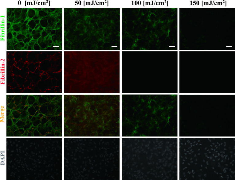
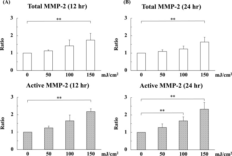
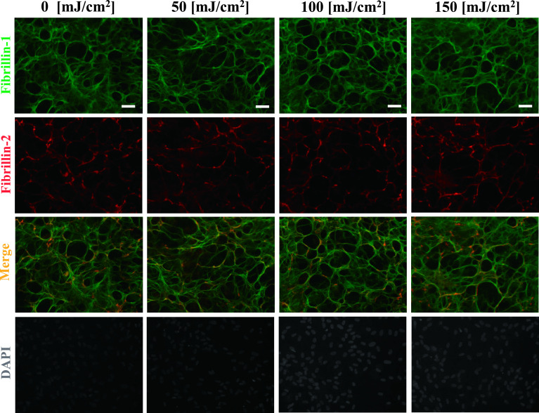
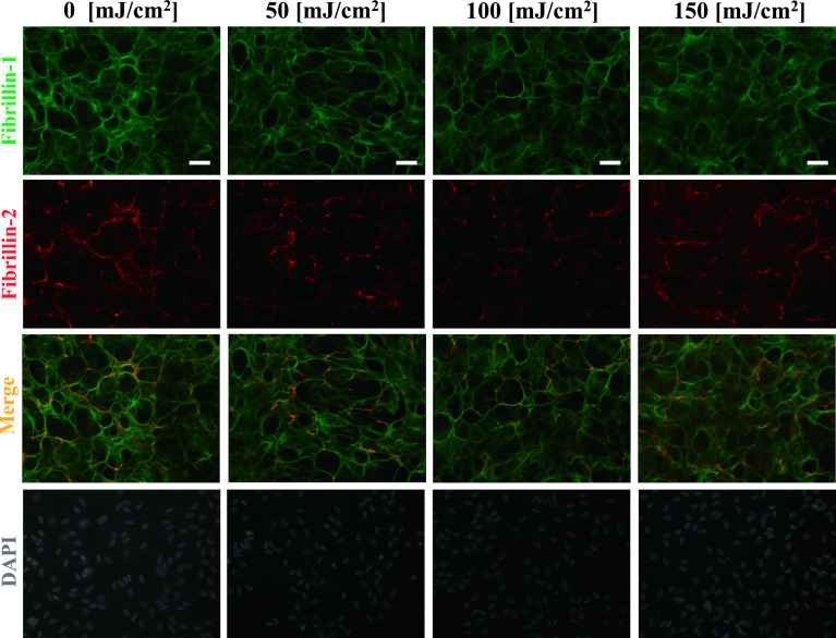
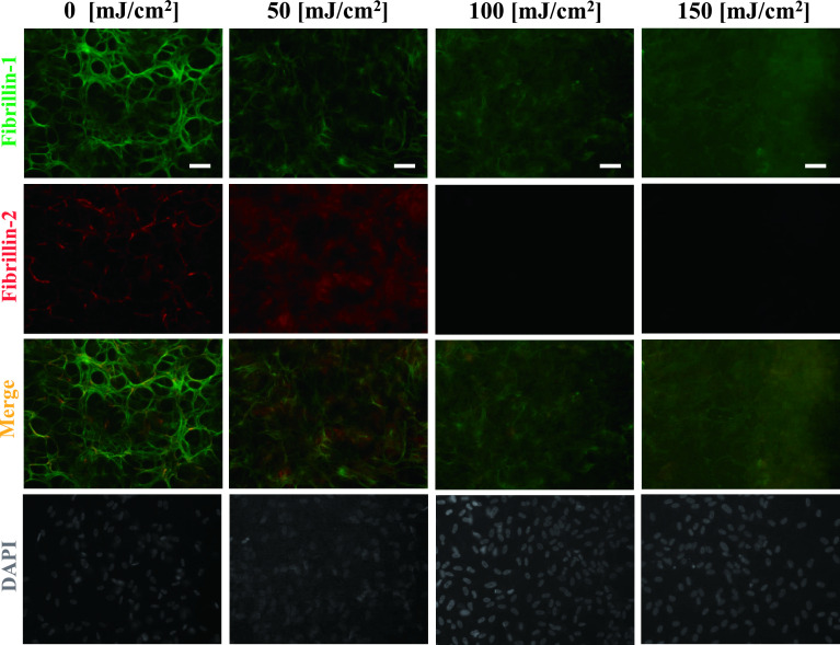
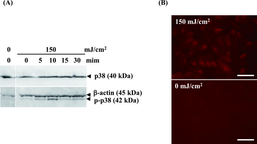
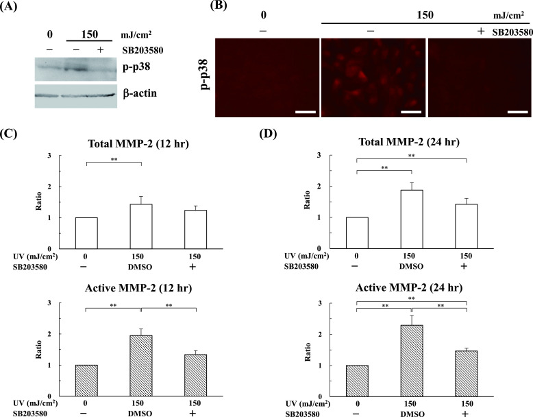
The ciliary zonules, also known as the zonules of Zinn, help to control the thickness of the lens during focusing. The ciliary zonules are composed of oxytalan fibers, which are synthesized by human nonpigmented ciliary epithelial cells (HNPCEC). The ciliary zonules are exposed to ultraviolet (UV), especially UV-A and UV-B, throughout life. We previously demonstrated that UV-B, but not UV-A, degrades fibrillin-1- and fibrillin-2-positive oxytalan fibers. However, the mechanism by which UV-B degrades oxytalan fibers remains unknown. In this study, we investigate the involvement of matrix metalloproteinase-2 (MMP-2) in the UV-B-induced degradation of fibrillin-1- and fibrillin-2-positive oxytalan fibers in cultured HNPCECs. Enzyme-linked immunosorbent assay revealed that UV-B irradiation at levels of 100 and 150 mJ/cm2 significantly increased the level of active MMP-2. Notably, MMP-2 inhibitors completely suppressed the degradation of fibrillin-1- and fibrillin-2-positive oxytalan fibers. In addition, we show that UV-B activates MMP-2 via stress-responsive kinase p38. Taken together, the results suggest that UV-B activates a production of active type of MMP-2 via the p38 pathway, and subsequently, an active-type MMP-2 degrades the fibrillin-1- and fibrillin-2-positive oxytalan fibers in cultured HNPCECs.
Keywords: ciliary zonule; fibrillin; matrix metalloproteinase-2; p38; ultraviolet-B.
2021 The Japan Society of Histochemistry and Cytochemistry.
Conflict of interest statement
VThe authors declare that there are no conflicts of interest.
Figures
Similar articles
-
The Effect of Ultraviolet B on Fibrillin-1 and Fibrillin-2 in Human Non-pigmented Ciliary Epithelial Cells In Vitro.Acta Histochem Cytochem. 2017 Jun 26;50(3):105-109. doi: 10.1267/ahc.16036. Epub 2017 Jun 14. Acta Histochem Cytochem. 2017. PMID: 28744027 Free PMC article.
-
Matrix metalloproteinase-2 degrades fibrillin-1 and fibrillin-2 of oxytalan fibers in the human eye and periodontal ligaments in vitro.Acta Histochem Cytochem. 2013 Oct 30;46(5):153-9. doi: 10.1267/ahc.13024. Epub 2013 Oct 23. Acta Histochem Cytochem. 2013. PMID: 24194629 Free PMC article.
-
Fibrillin-1 and fibrillin-2 are essential for formation of thick oxytalan fibers in human nonpigmented ciliary epithelial cells in vitro.Connect Tissue Res. 2012;53(1):14-20. doi: 10.3109/03008207.2011.602767. Epub 2011 Aug 18. Connect Tissue Res. 2012. PMID: 21851253
-
Corneal stroma microfibrils.Exp Eye Res. 2015 Mar;132:198-207. doi: 10.1016/j.exer.2015.01.014. Epub 2015 Jan 19. Exp Eye Res. 2015. PMID: 25613072 Free PMC article. Review.
-
Fibrillin microfibrils.Adv Protein Chem. 2005;70:405-36. doi: 10.1016/S0065-3233(05)70012-7. Adv Protein Chem. 2005. PMID: 15837522 Review.
Cited by
-
An Advanced Detection System for In Situ Hybridization Using a Fluorescence Resonance Energy Transfer-based Molecular Beacon Probe.Acta Histochem Cytochem. 2022 Oct 28;55(5):119-128. doi: 10.1267/ahc.22-00075. Epub 2022 Oct 25. Acta Histochem Cytochem. 2022. PMID: 36405552 Free PMC article.
-
A Novel Multi-Observation System to Study the Effects of Anterior Ocular Inflammation in Zinn's Zonule Using One Specimen.Int J Mol Sci. 2023 Mar 26;24(7):6254. doi: 10.3390/ijms24076254. Int J Mol Sci. 2023. PMID: 37047225 Free PMC article.
-
Morphologic features of crystalline lens in cataract patients with different lens sclerosis and axial length.BMC Ophthalmol. 2025 Jul 3;25(1):391. doi: 10.1186/s12886-025-04148-y. BMC Ophthalmol. 2025. PMID: 40610951 Free PMC article.
References
-
- Ashworth, J. L., Kelly, V., Rock, M. J., Shuttleworth, C. A. and Kielty, C. M. (1999) Regulation of fibrillin carboxy-terminal furin processing by N-glycosylation, and association of amino- and carboxy-terminal sequences. J. Cell Sci. 112; 4163–4171. - PubMed
-
- Fullmer, H. M. and Lillie, R. D. (1958) The oxytalan fiber: a previously undescribed connective tissue fiber. J. Histochem. Cytochem. 6; 425–430. - PubMed
LinkOut - more resources
Full Text Sources
Other Literature Sources
Miscellaneous