. 2021 Feb 26:12:637654.
doi: 10.3389/fimmu.2021.637654. eCollection 2021.

A Novel DNA Vaccine Against SARS-CoV-2 Encoding a Chimeric Protein of Its Receptor-Binding Domain (RBD) Fused to the Amino-Terminal Region of Hepatitis B Virus preS1 With a W4P Mutation

Affiliations

A Novel DNA Vaccine Against SARS-CoV-2 Encoding a Chimeric Protein of Its Receptor-Binding Domain (RBD) Fused to the Amino-Terminal Region of Hepatitis B Virus preS1 With a W4P Mutation

Hyein Jeong et al. Front Immunol. .

Abstract

A coronavirus SARS-CoV-2, which has caused the pandemic viral pneumonia disease COVID-19, significantly threatens global public health, highlighting the need to develop effective and safe vaccines against its infection. In this study, we developed a novel DNA vaccine candidate against SARS-CoV-2 by expressing a chimeric protein of its receptor-binding domain (RBD) fused to a 33-bp sequence (11 aa) from the hepatitis B virus (HBV) preS1 region with a W4P mutation (W4P-RBD) at the N-terminal region and evaluated its immunogenicity. In vitro transfection experiments in multiple cell lines demonstrated that W4P-RBD vs. wild-type RBD protein (W-RBD) led to enhanced production of IL-6 and TNFα at the transcription and translation levels, suggesting the adjuvant potential of N-terminal HBV preS1 sequences for DNA vaccines against SARS-CoV-2. W4P-RBD also led to enhanced production of IgG and IgA, which can neutralize and block SARS-CoV-2 infection in both blood sera and bronchoalveolar lavage (BAL) fluid from the lung in vaccinated mice. Additionally, W4P-RBD led to an enhanced T-cell-mediated cellular immune response under S1 protein stimulation. In summary, W4P-RBD led to robust humoral and cell-mediated immune responses against SARS-CoV-2 in vaccinated mice, highlighting its feasibility as a novel DNA vaccine to protect against SARS-CoV-2 infection.

Keywords: COVID-19; DNA vaccine; HBV preS1; Receptor-binding domain (RBD); SARS-CoV-2; W4P-RBD.


Conflict of interest statement

The authors declare that the research was conducted in the absence of any commercial or financial relationships that could be construed as a potential conflict of interest.

Figures

Figure 1
Figure 1
Construction of the HBV W4P preS1-fused pcDNA3.3-RBD plasmid (W4P-RBD) as a candidate for SARS-CoV-2. (A) Design of pcDNA3.3-RBD and pcDNA3.3-W4P-RBD. The W4P region comprises 33 bp from the first site of the preS1 region of the HBV genome and encodes 11 amino acids. (B) The protein expression of SARS-CoV-2 RBD and W4P-conjugated RBD was detected by the Western blot assay. pcDNA3.3-RBD, pcDNA3.3-W4P-RBD, and empty pcDNA3.3 were transfected into Vero E6, Huh7, and 293T cells, and cell lysates were collected 48 h post transfection to detect protein expression. (C) The mRNA expression levels of IL-6 and in pcDNA3.3-transfected cells were detected by qRT-PCR. Significance differences (*P < 0.05, **P < 0.01, ***P < 0.001) among the different groups are shown in the related figures, and the data are presented as the means ± s.e.m. of three independent experiments.
Figure 2
Figure 2
W4P-RBD elicits RBD-specific antibody responses in serum and induces potent neutralizing activity against pseudotyped SARS-CoV-2. C57BL/6 mice were immunized with W-RBD, W4P-RBD (50 μg/mouse), or empty pcDNA3.3 (Mock) three times at 1-week intervals. (A,C) Antibody responses in serum specific to SARS-CoV-2 RBD proteins were detected by ELISA. (B) Serum at 5 weeks after the last immunization was assessed using different dilution factors for IgG against the SARS-CoV-2 RBD protein using ELISA. (D) The 50% neutralizing antibody titer (NT50) was calculated using the SARS-CoV-2 pseudovirus neutralization assay in Calu-3 cells. (E) Correlation between SARS-CoV-2 RBD-specific IgG and pseudotyped SARS-CoV-2 neutralization titers for immunized mice. Significance differences (*P < 0.05, **P < 0.01, ***P < 0.001) among the different groups are shown in the related figures, and the data are presented as the means ± s.e.m. of mice (n = 7). Pearson's correlations were calculated to define correlations.
Figure 3
Figure 3
W4P-RBD exerts potent neutralizing activity against live SARS-CoV-2. C57BL/6 mice were immunized with W-RBD, W4P-RBD (50 μg/mouse), or empty pcDNA3.3 (Mock) three times at 1-week intervals. Serum from the immunized mice was diluted and incubated with live SARS-CoV-2 for neutralization assays. (A) A 50% plaque reduction neutralizing antibody (PRNT50) titer against live SARS-CoV-2 was calculated against SARS-CoV-2 infection in Vero E6 cells. (B) Reduction in plaque formation in Vero E6 cells infected with SARS-CoV-2. (C) Correlation between SARS-CoV-2 RBD-specific IgG and SARS-CoV-2 neutralization titers in immunized mice. Significance differences (**P < 0.01, ***P < 0.001) among the different groups are shown in the related figures, and the data are presented as the means ± s.e.m. of mice (n = 7). Pearson's correlations were calculated to define correlations.
Figure 4
Figure 4
The W4P-RBD vaccine exerts potent neutralizing activity against live SARS-CoV-2 and inhibits viral infection and replication of SARS-CoV-2. C57BL/6 mice were immunized with W-RBD, W4P-RBD (50 μg/mouse), or empty pcDNA3.3 (Mock) three times at 1-week intervals. Serum from the immunized mice was diluted and incubated with live SARS-CoV-2 for neutralization assays. (A) The neutralization efficacy of serum from immunized mice against live SARS-CoV-2 RNA copy number in Vero E6 cells was determined by qRT-PCR. (B) The neutralization efficacy of the diluted serum against live SARS-CoV-2 in Vero E6 cells was determined by Western blotting. (C) Pooled serum (diluted 1:500) from each mouse group was tested in the neutralization assay against live SARS-CoV-2. Representative images (40-fold magnification) show live SARS-CoV-2-infected cells after neutralization in each group. Significance differences (*P < 0.05) among the different groups are shown in the related figures, and the RNA data are presented as the means ± s.e.m. of mice (n = 7).
Figure 5
Figure 5
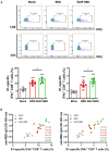
W4P-RBD potentiates functional T cells specific to SARS-CoV-2 S1 proteins. C57BL/6 mice were intramuscularly injected with W-RBD, W4P-RBD (50 μg/mouse), or mock, and the spleens were collected 5 weeks post-vaccination for analysis by flow cytometry. (A,B) Splenocytes were incubated with SARS-CoV-2 S1 protein (5 μg/ml) for 24 h and stained to detect IFNγ-producing CD8+ T cells and CD4+ T cells. (C) Correlation between RBD-specific IgG in serum and the S1-specific T-cell population in splenocytes. Significance differences (*P < 0.05, ***P < 0.001) among the different groups are shown in the related figures, and the data are presented as the means ± s.e.m. of mice (n = 7). Pearson's correlations were calculated to define correlations.
Figure 6
Figure 6
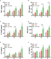
W4P-RBD induces proinflammatory cytokine production in S1-stimulated splenocytes. Splenocytes from immunized mice were stimulated with SARS-CoV-2 S1 protein (5 μg/ml) for 5 days. The cytokine production of (A) TNFα, (B) IFNγ, (C) IL-12p40, (D) IFNβ, (E) IL-6, and (F) IL-2 from splenocytes was detected by ELISA. Significance differences (*P < 0.05, **P < 0.01, ***P < 0.001) among the different groups are shown in the related figures, and the data are presented as the means ± s.e.m. of mice (n = 7).
Figure 7
Figure 7
Schematic representation of W4P-RBD as a vaccine candidate against SARS-CoV-2. A novel platform of N-terminal addition of HBV W4P preS1 33-bp sequences for a DNA vaccine against SARS-CoV-2 were developed. The W4P-RBD led to enhanced both humoral and cell-mediated immune response against SARS-CoV-2 in vaccinated mice, demonstrating its feasibility as a DNA vaccine to protect against SARS-CoV-2.

Similar articles

Cited by

References

    1. Chan J, Kok K, Zhu Z, To K, Yuan S, Yuen K, et al. . Genomic characterization of the 2019 novel human pathogenic coronavirus isolated from a patient with atypical pneumonia after visiting Wuhan. Emerg Microbes Infect. (2020) 9:221–36. 10.1080/22221751.2020.1719902 - DOI - PMC - PubMed
    1. Huang C, Wang Y, Li X, Ren L, Zhao J, Hu Y, et al. . Clinical features of patients infected with 2019 novel coronavirus in Wuhan, China. Lancet. (2020) 395:497–506. 10.1016/S0140-6736(20)30183-5 - DOI - PMC - PubMed
    1. Li H, Liu L, Zhang D, Xu J, Dai H, Tang N, et al. . SARS-CoV-2 and viral sepsis: observations and hypotheses. Lancet. (2020) 395:1517–20. 10.1016/S0140-6736(20)30920-X - DOI - PMC - PubMed
    1. Wu Y, Wang F, Shen C, Peng W, Li D, Zhao C, et al. . A noncompeting pair of human neutralizing antibodies block COVID-19 virus binding to its receptor ACE2. Science. (2020) 368:1274–8. 10.1126/science.abc2241 - DOI - PMC - PubMed
    1. Zhu N, Zhang D, Wang W, Li X, Yang B, Song J, et al. . A novel coronavirus from patients with pneumonia in China. N Engl J Med. (2020) 382:727–33. 10.1056/NEJMoa2001017 - DOI - PMC - PubMed

Publication types

MeSH terms