

Salvianolic acid B protects against acute and chronic liver injury by inhibiting Smad2C/L phosphorylation
- PMID: 33732314
- PMCID: PMC7903446
- DOI: 10.3892/etm.2021.9772
Salvianolic acid B protects against acute and chronic liver injury by inhibiting Smad2C/L phosphorylation
Abstract
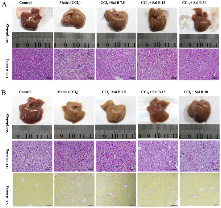
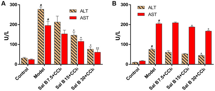
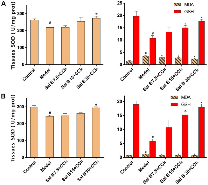
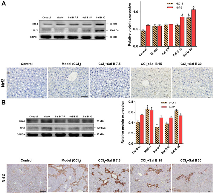
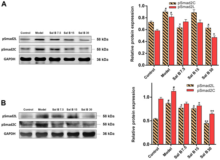
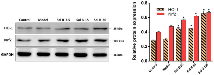
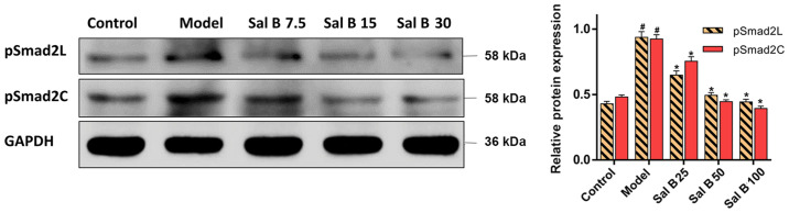
Salvianolic acid B (Sal B) has strong antioxidant and anti-fibrosis effects, which are related to the transforming growth factor β/Smad signaling pathway. However, how Sal B affects this antioxidant pathway and the phosphorylation (p-) of Smad2 at both the COOH-terminal (pSmad2C) and linker region (pSmad2L) are unknown. The aims of the present study were to investigate the underlying mechanisms of Sal B on acute and chronic liver injury induced by CCl4 and H2O2, and its effects on p-Smad2C/L. In in vivo experiments, acute and chronic liver injury models were induced by CCl4, and the oxidative damage cell model was established in vitro with H2O2. Liver histopathology was assessed using hematoxylin and eosin and Van Gieson's staining. Moreover, serum biochemical indicators were analyzed using specific assay kits. Furthermore, the present study evaluated the oxidant/antioxidant status in acute and chronic liver injury models by oxidative stress parameters such as malondialdehyde, glutathione and superoxide dismutase. In addition, western blot analysis was performed to analyze the protein expression levels of pSmad2C, pSmad2L, nuclear factor erythroid-2-related factor 2 (Nrf2) and heme oxygenase-1 (HO-1). It was found that Sal B improved liver histology, decreased the levels of aminotransferase and attenuated oxidative stress in acute and chronic liver injury models. Additionally, the protein expression levels of pSmad2C and pSmad2L were decreased, but Nrf2 and HO-1 expression levels were increased both in vivo and in vitro. Collectively, the present results suggested that Sal B may protect against acute and chronic liver injury via inhibition of Smad2C/L phosphorylation, and the Nrf2/HO-1 signaling pathway may play an important role in this process.
Keywords: liver injury; nuclear factor erythroid-2-related factor 2/heme oxygenase-1; phosphorylated Smad2 at COOH-terminal/linker region; salvianolic acid B.
Copyright: © Tao et al.
Conflict of interest statement
The authors declare that they have no competing interests.
Figures
Similar articles
-
Salvianolic acid B exerts a protective effect in acute liver injury by regulating the Nrf2/HO-1 signaling pathway.Can J Physiol Pharmacol. 2020 Mar;98(3):162-168. doi: 10.1139/cjpp-2019-0349. Epub 2019 Oct 11. Can J Physiol Pharmacol. 2020. PMID: 31604020
-
Salvianolic acid B exerts anti-liver fibrosis effects via inhibition of MAPK-mediated phospho-Smad2/3 at linker regions in vivo and in vitro.Life Sci. 2019 Dec 15;239:116881. doi: 10.1016/j.lfs.2019.116881. Epub 2019 Oct 31. Life Sci. 2019. PMID: 31678285
-
Combination of chlorogenic acid and salvianolic acid B protects against polychlorinated biphenyls-induced oxidative stress through Nrf2.Environ Toxicol Pharmacol. 2016 Sep;46:255-263. doi: 10.1016/j.etap.2016.08.004. Epub 2016 Aug 3. Environ Toxicol Pharmacol. 2016. PMID: 27513569
-
Activation of Nrf2/AREs-mediated antioxidant signalling, and suppression of profibrotic TGF-β1/Smad3 pathway: a promising therapeutic strategy for hepatic fibrosis - A review.Life Sci. 2020 Sep 1;256:117909. doi: 10.1016/j.lfs.2020.117909. Epub 2020 Jun 5. Life Sci. 2020. PMID: 32512009 Review.
-
Role of salvianolic acid B in the treatment of acute ischemic stroke: a systematic review and meta-analysis of animal models.Front Pharmacol. 2024 Dec 24;15:1479765. doi: 10.3389/fphar.2024.1479765. eCollection 2024. Front Pharmacol. 2024. PMID: 39776581 Free PMC article. Review.
Cited by
-
Salvianolic acid B suppresses hepatic fibrosis by inhibiting ceramide glucosyltransferase in hepatic stellate cells.Acta Pharmacol Sin. 2023 Jun;44(6):1191-1205. doi: 10.1038/s41401-022-01044-9. Epub 2023 Jan 10. Acta Pharmacol Sin. 2023. PMID: 36627345 Free PMC article.
-
Metabolic Reprograming of Macrophages: A New Direction in Traditional Chinese Medicine for Treating Liver Failure.J Immunol Res. 2024 Dec 24;2024:5891381. doi: 10.1155/jimr/5891381. eCollection 2024. J Immunol Res. 2024. PMID: 39741958 Free PMC article. Review.
-
MST1/2 DKO abates salvianolic acid B's therapeutic effect on CCl4-induced liver injury mice.Naunyn Schmiedebergs Arch Pharmacol. 2025 Apr 12. doi: 10.1007/s00210-025-04140-9. Online ahead of print. Naunyn Schmiedebergs Arch Pharmacol. 2025. PMID: 40220025
-
A Comprehensive Review of Rosmarinic Acid: From Phytochemistry to Pharmacology and Its New Insight.Molecules. 2022 May 20;27(10):3292. doi: 10.3390/molecules27103292. Molecules. 2022. PMID: 35630768 Free PMC article. Review.
-
Salvianolic Acid B: A Review of Pharmacological Effects, Safety, Combination Therapy, New Dosage Forms, and Novel Drug Delivery Routes.Pharmaceutics. 2023 Aug 29;15(9):2235. doi: 10.3390/pharmaceutics15092235. Pharmaceutics. 2023. PMID: 37765204 Free PMC article. Review.
References
-
- Ramadori G, Moriconi F, Malik I, Dudas J. Physiology and pathophysiology of liver inflammation, damage and repair. J Physiol Pharmacol. 2008;59:107–117. - PubMed
LinkOut - more resources
Full Text Sources
Other Literature Sources