. 2021 Apr;21(4):286.
doi: 10.3892/ol.2021.12547. Epub 2021 Feb 12.

Curcumin inhibits the growth of liver cancer by impairing myeloid-derived suppressor cells in murine tumor tissues

Affiliations

Curcumin inhibits the growth of liver cancer by impairing myeloid-derived suppressor cells in murine tumor tissues

Sha Tian et al. Oncol Lett. 2021 Apr.

Abstract

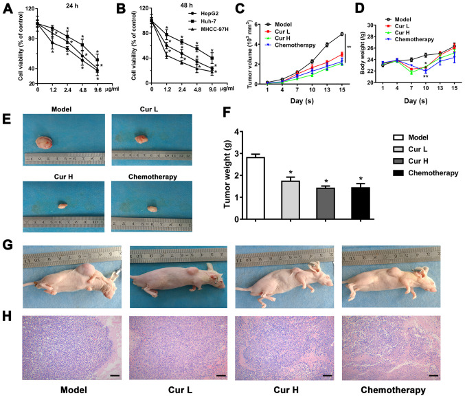
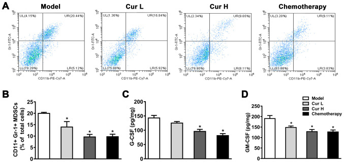
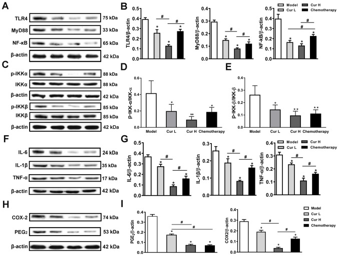
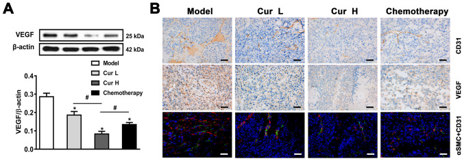
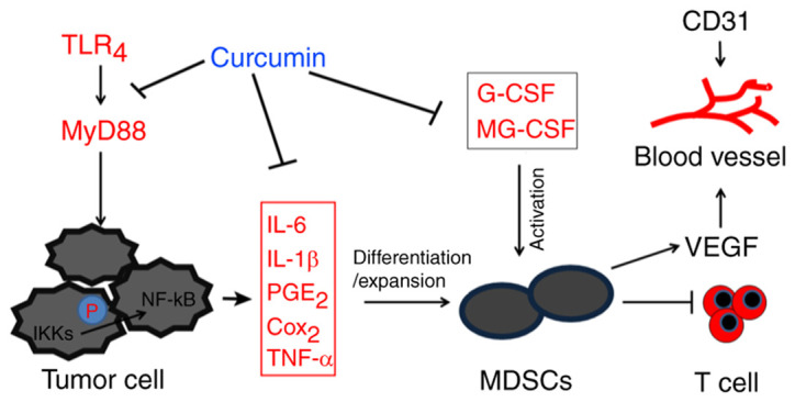
Curcumin, one of the active ingredients of Curcuma longa (Jianghuang), has been reported to exert multiple bioactivities, including pro-apoptotic and anti-inflammatory activities. In recent years, curcumin has been extensively studied, and it has been revealed that curcumin inhibits the growth of numerous types of cancer. However, to the best of our knowledge, the inhibitory effects of curcumin on the activation or expansion of myeloid-derived suppressor cells (MDSCs) in liver cancer and the underlying mechanism have not yet been determined. Therefore, the present study aimed to investigate the inhibitory effect of curcumin on MDSC activity and the associated anti-neoplastic mechanism in a HepG2 ×enograft mouse model. The effect of curcumin on the viability of Huh-7, MHCC-97H and HepG2 cells in vitro was analyzed using a Cell Counting Kit-8 assay. The effects of curcumin on tumor growth, numbers of MDSCs, expression levels of proteins involved in the toll-like receptor 4 (TLR4)/NF-κB signaling pathway, levels of related inflammatory factors and angiogenesis were determined in HepG2 ×enograft model mice, which were given different doses of curcumin via intragastrical administration. The results of the present study revealed that curcumin inhibited the viability of Huh-7, MHCC-97H and HepG2 cells and the growth of HepG2 ×enograft tumors in mice. Flow cytometric analysis indicated that curcumin reduced the number of MDSCs in mouse xenograft tumors. In addition, the results demonstrated that curcumin inhibited the TLR4/NF-κB signaling pathway and the expression of inflammatory factors, including IL-6, IL-1β, prostaglandin E2 and cyclooxygenase-2, in mouse xenograft tumors. Furthermore, curcumin suppressed the secretion of granulocyte-macrophage colony-stimulating factor (GM-CSF) and granulocyte-colony stimulating factor (G-CSF), which are essential factors for MDSCs modulation, in tumor tissues. Additionally, curcumin was revealed to inhibit angiogenesis, which was demonstrated by the downregulation of the expression levels of vascular endothelial growth factor, CD31 and α-smooth muscle actin in western blotting, immunohistochemistry and immunofluorescence experiments. In conclusion, the findings of the present study identified a novel mechanism via which curcumin may suppress the growth of liver cancer by reducing the numbers of MDSCs and subsequently disrupting the process of angiogenesis. These conclusions were supported by the observed inactivation of the TLR4/NF-κB signaling pathway-mediated inflammatory response and the downregulation of GM-CSF and G-CSF secretion in xenograft tissues.

Keywords: curcumin; liver cancer; myeloid-derived suppressor cells; toll-like receptor 4/NF-κB; vascular endothelial growth factor.


Conflict of interest statement

The authors declare that they have no competing interests.

Figures

Figure 1.
Figure 1.
Curcumin suppresses liver cancer growth in vivo and in vitro. Viability of HepG2, Huh-7 and MHCC-97H cells following curcumin treatment at (A) 24 and (B) 48 h. The x-axis presents the concentration of curcumin (µg/ml). Effects of curcumin on (C) tumor volume and (D) body weight were measured every 3 days. (E) Representative images of excised tumors in each group. (F) Weight of tumor tissues on day 15. (G) Representative images of nude mice with tumor xenografts in each group. (H) Hematoxylin and eosin staining of tumor tissues (magnification, ×100; scale bar, 200 µm). Data are presented as the mean ± SD (n=7). *P<0.05 and **P<0.01 vs. model group or vehicle control in CCK-8 assay. Cur L, curcumin low dose group (120 mg/kg); Cur H, curcumin high dose group (240 mg/kg).
Figure 2.
Figure 2.
Curcumin inhibits MDSCs in tumor tissues. (A) Flow cytometric analysis of CD11b+/Gr-1+ cells in murine tumor tissues. (B) Quantitative analysis of MDSCs in murine tumor tissue. Secretory levels of (C) G-CSF and (D) GM-CSF in the tumor tissues were measured using ELISAs. Tumor tissue samples were collected within 24 h following the attenuation of treatment with all drugs. Data are presented as the mean ± SD (n=7). *P<0.05 vs. model group. MDSCs, myeloid-derived suppressor cells; GM-CSF, granulocyte-macrophage colony-stimulating factor; G-CSF, granulocyte-colony-stimulating factor; Cur L, curcumin low dose group (120 mg/kg); Cur H, curcumin high dose group (240 mg/kg); Gr-1, granulocyte receptor-1; UL, upper left; UR, upper right; LL, lower left; LR, lower right.
Figure 3.
Figure 3.
Inhibition of the TLR4/NF-κB signaling pathway-mediated inflammatory response. (A) Western blotting was used to analyze and (B) semi-quantify the expression levels of TLR4, MyD88 and NF-κB in murine tumor tissues. (C) Expression levels and semi-quantitative analysis of (D) IKK-α, p-IKK-α, (E) IKK-β and p-IKK-β in murine tumor tissues. (F) Expression levels and (G) semi-quantitative analysis of IL-6, IL-1β and TNF-α in murine tumor tissues. (H) Expression levels and (I) semi-quantitative analysis of PGE2 and COX-2 in murine tumor tissues. Data are presented as the mean ± SD (n=7). *P<0.05 and **P<0.01 vs. model group; #P<0.05, as indicated. TLR4, toll-like receptor 4; MyD88, myeloid differentiation primary response 88; IKKα, nuclear factor-κB kinase α; IKKβ, nuclear factor-κB kinase β; PGE2, prostaglandin E2; COX-2, cyclooxygenase-2; p-, phosphorylated; Cur L, curcumin low dose group (120 mg/kg); Cur H, curcumin high dose group (240 mg/kg).
Figure 4.
Figure 4.
Curcumin suppresses tumor angiogenesis. (A) Western blotting was used to analyze and semi-quantify the expression levels of VEGF. (B) Immunohistochemistry analysis of CD31 and VEGF expression in murine tumor tissues, and immunofluorescence analysis of αSMC and CD31 expression in murine tumor tissues (magnification, ×400; scale bar, 50 µm). Data are presented as the mean ± SD (n=7). *P<0.05 vs. model group; #P<0.05, as indicated. VEGF, vascular endothelial growth factor; αSMC, α-smooth muscle cell actin; Cur L, curcumin low dose group (120 mg/kg); Cur H, curcumin high dose group (240 mg/kg).
Figure 5.
Figure 5.
Schematic diagram of the inhibitory effect of curcumin on MDSCs and liver cancer. Curcumin exerted an inhibitory effect on MDSCs by inhibiting the TLR4/NF-κB-mediated inflammatory microenvironment and attenuating G-CSF and GM-CSF secretion. This subsequently suppresses tumor angiogenesis and ameliorates immune tolerance in liver cancer, thus exhibiting anti-liver cancer effects. TLR4, toll-like receptor 4; MyD88, myeloid differentiation primary response 88; IKK, nuclear factor-κB kinase; PGE2, prostaglandin E2; COX-2, cyclooxygenase-2; MDSCs, myeloid-derived suppressor cells; G-CSF, granulocyte-colony stimulating factor; GM-CSF, granulocyte-macrophage colony-stimulating factor; VEGF, vascular endothelial growth factor.

Similar articles

Cited by

References

    1. Galon J, Costes A, Sanchez-Cabo F, Kirilovsky A, Mlecnik B, Lagorce-Pagès C, Tosolini M, Camus M, Berger A, Wind P, et al. Type, density, and location of immune cells within human colorectal tumors predict clinical outcome. Science. 2006;313:1960–1964. doi: 10.1126/science.1129139. - DOI - PubMed
    1. Condeelis J, Pollard JW. Macrophages: Obligate partners for tumor cell migration, invasion, and metastasis. Cell. 2006;124:263–266. doi: 10.1016/j.cell.2006.01.007. - DOI - PubMed
    1. Movahedi K, Guilliams M, Van den Bossche J, Van den Bergh R, Gysemans C, Beschin A, De Baetselier P, Van Ginderachter JA. Identification of discrete tumor-induced myeloid-derived suppressor cell subpopulations with distinct T cell-suppressive activity. Blood. 2008;111:4233–4244. doi: 10.1182/blood-2007-07-099226. - DOI - PubMed
    1. De Sanctis F, Solito S, Ugel S, Molon B, Bronte V, Marigo I. MDSCs in cancer: Conceiving new prognostic and therapeutic targets. Biochim Biophys Acta. 2016;1865:35–48. - PubMed
    1. Watson GA, Fu YX, Lopez DM. Splenic macrophages from tumor-bearing mice co-expressing MAC-1 and MAC-2 antigens exert immunoregulatory functions via two distinct mechanisms. J Leukoc Biol. 1991;49:126–138. doi: 10.1002/jlb.49.2.126. - DOI - PubMed