Case Reports
. 2021 Mar 8;2021(3):omab003.
doi: 10.1093/omcr/omab003. eCollection 2021 Mar.

Evaluation and management of a spontaneous corneal rupture secondary to pellucid marginal degeneration, using swept-source anterior segment optical coherence tomography

Affiliations
Case Reports

Evaluation and management of a spontaneous corneal rupture secondary to pellucid marginal degeneration, using swept-source anterior segment optical coherence tomography

Esther Papamlichael et al. Oxf Med Case Reports. .

Abstract

We describe a case of bilateral spontaneous corneal perforation secondary to pellucid marginal degeneration and present the associated swept-source anterior segment optical coherence tomography (SS-ASOCT) findings and management principles used. A 47-year-old woman presented with ocular pain, redness, foreign body sensation and clear discharge in the right eye in 2017 and with very similar symptoms in 2019 in the left eye. Clinically she had a corneal perforation at the inferior cornea with associated loss of anterior chamber volume. Corneal topography demonstrated peripheral thinning and steepening in the contralateral eye. ASOCT images revealed full-thickness perforation, iridocorneal touch and iris stranding. The patient was managed with a combination of contact bandaging and corneal gluing. SS-ASOCT is a useful adjunctive tool in the clinical assessment and evaluation of spontaneous corneal perforation. Alongside the clinical evaluation, it can be used to monitor the clinical response.


Figures

Figure 1
Figure 1
Inferiotemporal peripheral cornea with a bandage contact lens in situ (A) and associated localized edema (B). SS-ASOCT image revealed the presence of an oblique corneal perforation and localized corneal edema (C, D) as well as iridocorneal touch near the site of perforation (D).
Figure 2
Figure 2
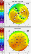
Pentacam® (Oculus) corneal topography of the contralateral eye demonstrating evidence of peripheral steepening and corneal thinning consistent with a diagnosis of PMD.
Figure 3
Figure 3
Seven-week follow-up SS-ASOCT images showing partial resolution of the corneal perforation in the posterior stroma with remaining disorganization anteriorly. Peripheral thinning is also demonstrated in these images consistent with the diagnosis of PMD.
Figure 4
Figure 4
Nine-week anterior segment follow-up pictures showing that the perforation healed (A) with no leakage and no residual epithelial deficit (B). This was confirmed with SS-ASCOT showing the formation of stromal scar in the area previously perforated and localized thinning (C).

References

    1. Sridhar MS, Mahesh S, Bansal AK, Nutheti R, Rao GN. Pellucid marginal corneal degeneration. Ophthalmology 2004;111:1102–7. doi: 10.1016/j.ophtha.2003.09.035. - DOI - PubMed
    1. Basu S, Vaddavalli PK, Vemuganti GK, Md Hasnat A, Somasheila IM. Anterior segment optical coherence tomography features of acute corneal hydrops. Cornea 2012;31:479–85. doi: 10.1097/ICO.0b013e318223988e. - DOI - PubMed
    1. Abdelazeem K, Sharaf M, Saleh MGA, Fathalla AM, Soliman W. Relevance of swept-source anterior segment optical coherence tomography for corneal imaging in patients with flap-related complications after LASIK. Cornea 2019;38:93–7. doi: 10.1097/ICO.0000000000001773. - DOI - PubMed
    1. Satue M, Idoipe M, Sanchez-Perez A, Liarakos VS, Mateo A, Garcia-Martin E, et al. Evaluation of early graft detachment after Descemet membrane endothelial keratoplasty using new swept-source optical coherence tomography. Cornea 2016;35:1279–84. doi: 10.1097/ICO.0000000000000925. - DOI - PubMed
    1. Steinberg J, Casagrande MK, Frings A, Katz T, Druchkiv V, Richard G, et al. Screening for subclinical keratoconus using swept-source Fourier domain anterior segment optical coherence tomography. Cornea 2015;34:1413–9. doi: 10.1097/ICO.0000000000000568. - DOI - PubMed

Publication types

LinkOut - more resources