

A comparative study of autogenous, allograft and artificial bone substitutes on bone regeneration and immunotoxicity in rat femur defect model
- PMID: 33732488
- PMCID: PMC7947581
- DOI: 10.1093/rb/rbaa040
A comparative study of autogenous, allograft and artificial bone substitutes on bone regeneration and immunotoxicity in rat femur defect model
Abstract
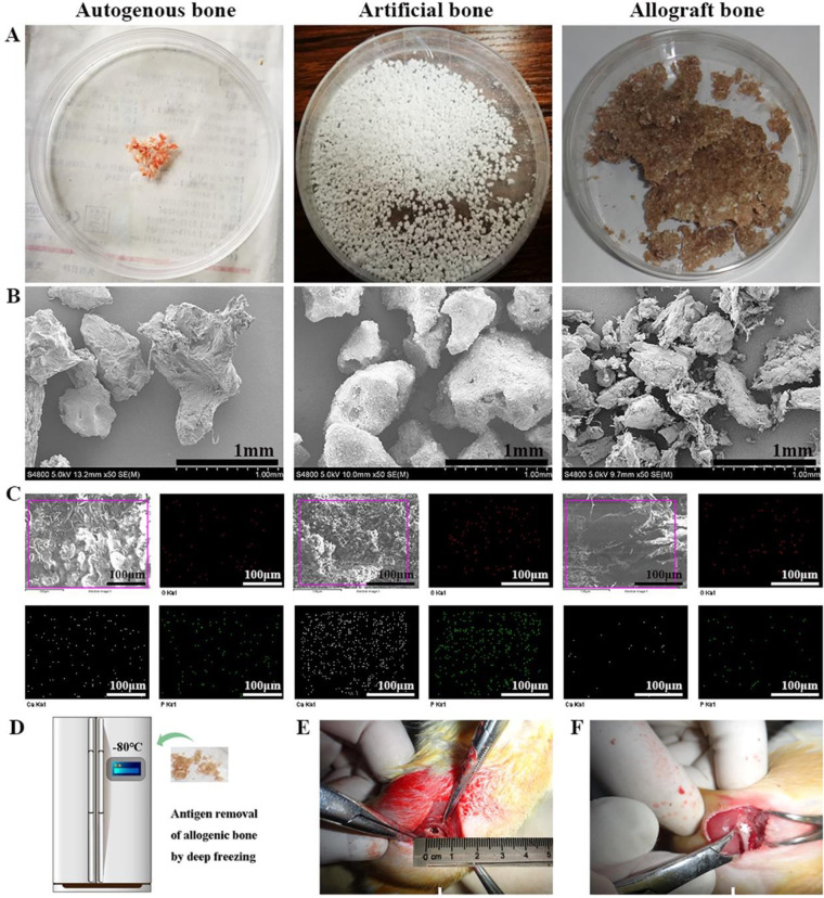
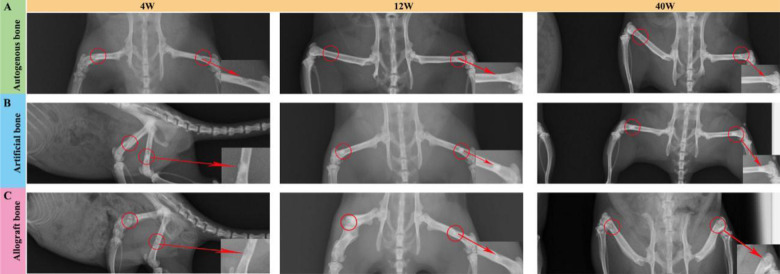
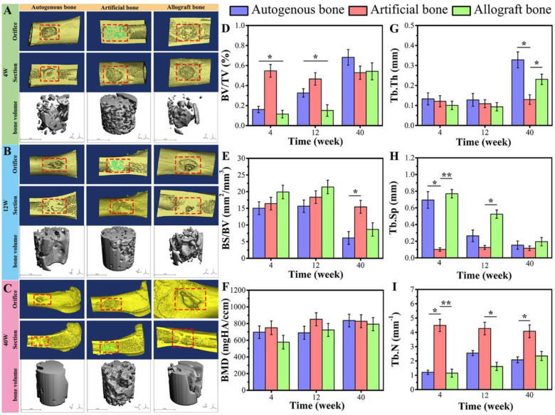
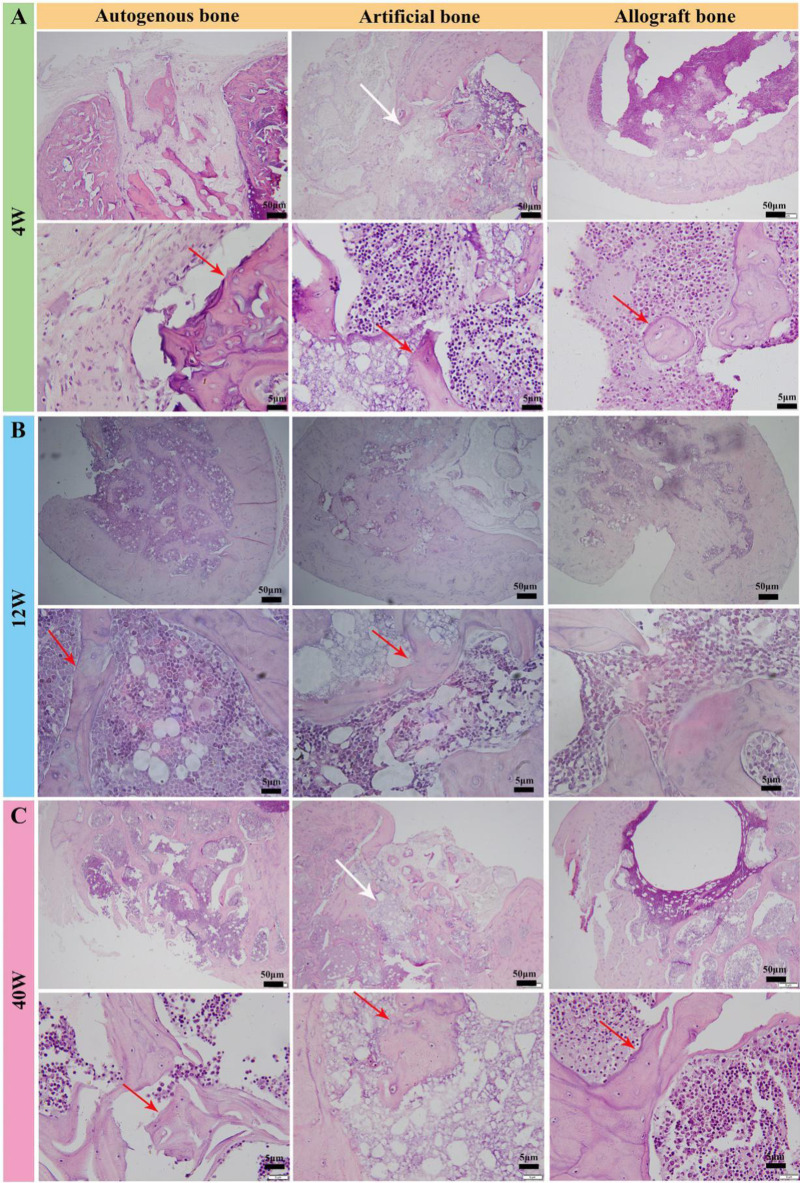
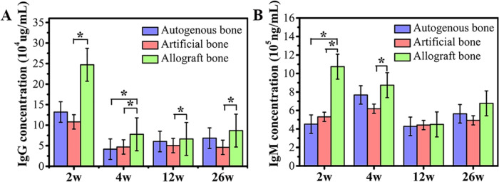
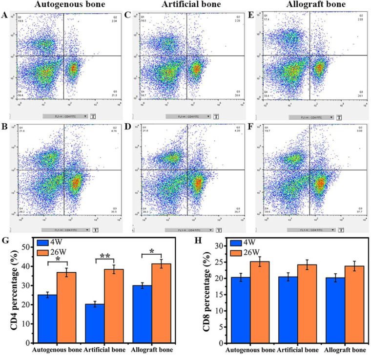
Repair and reconstruction of large bone defect were often difficult, and bone substitute materials, including autogenous bone, allogenic bone and artificial bone, were common treatment strategies. The key to elucidate the clinical effect of these bone repair materials was to study their osteogenic capacity and immunotoxicological compatibility. In this paper, the mechanical properties, micro-CT imaging analysis, digital image analysis and histological slice analysis of the three bone grafts were investigated and compared after different time points of implantation in rat femur defect model. Autogenous bone and biphasic calcium phosphate particular artificial bone containing 61.4% HA and 38.6% β-tricalcium phosphate with 61.64% porosity and 0.8617 ± 0.0068 g/cm3 density (d ≤ 2 mm) had similar and strong bone repair ability, but autogenous bone implant materials caused greater secondary damage to experimental animals; allogenic bone exhibited poor bone defect repair ability. At the early stage of implantation, the immunological indexes such as Immunoglobulin G, Immunoglobulin M concentration and CD4 cells' population of allogenic bone significantly increased in compared with those of autologous bone and artificial bone. Although the repair process of artificial bone was relatively inefficient than autologous bone graft, the low immunotoxicological indexes and acceptable therapeutic effects endowed it as an excellent alternative material to solve the problems with insufficient source and secondary trauma of autogenous bone.
Keywords: allogenic bone; artificial bone; autogenous bone; bone defect repair; immunotoxicity; osteogenesis.
© The Author(s) 2020. Published by Oxford University Press.
Figures

Similar articles
-
Evaluation of New Biphasic Calcium Phosphate Bone Substitute: Rabbit Femur Defect Model and Preliminary Clinical Results.J Med Biol Eng. 2017;37(1):85-93. doi: 10.1007/s40846-016-0203-3. Epub 2017 Jan 19. J Med Biol Eng. 2017. PMID: 28286465 Free PMC article.
-
Three-dimensional printing akermanite porous scaffolds for load-bearing bone defect repair: An investigation of osteogenic capability and mechanical evolution.J Biomater Appl. 2016 Nov;31(5):650-660. doi: 10.1177/0885328216664839. Epub 2016 Sep 1. J Biomater Appl. 2016. PMID: 27585972
-
Novel osteoconductive β-tricalcium phosphate/poly(L-lactide-co-e-caprolactone) scaffold for bone regeneration: a study in a rabbit calvarial defect.J Mater Sci Mater Med. 2018 Oct 8;29(10):156. doi: 10.1007/s10856-018-6159-9. J Mater Sci Mater Med. 2018. PMID: 30298429
-
Bone grafts and their substitutes.Bone Joint J. 2016 Jan;98-B(1 Suppl A):6-9. doi: 10.1302/0301-620X.98B.36350. Bone Joint J. 2016. PMID: 26733632 Review.
-
Ingrowth and formation of bone in defects in an uncemented fiber-metal total hip-replacement model in dogs.J Bone Joint Surg Am. 1991 Jan;73(1):93-105. J Bone Joint Surg Am. 1991. PMID: 1985999 Review.
Cited by
-
Surface demineralized freeze-dried bone allograft followed by reimplantation in a failed mandibular dental implant.Regen Biomater. 2023 Nov 17;11:rbad102. doi: 10.1093/rb/rbad102. eCollection 2024. Regen Biomater. 2023. PMID: 38173777 Free PMC article.
-
A sustained release of BMP2 in urine-derived stem cells enhances the osteogenic differentiation and the potential of bone regeneration.Regen Biomater. 2022 Apr 25;9:rbac015. doi: 10.1093/rb/rbac015. eCollection 2022. Regen Biomater. 2022. PMID: 35529046 Free PMC article.
-
3D printed scaffolds with multistage osteogenic activity for bone defect repair.Regen Biomater. 2025 Mar 10;12:rbaf010. doi: 10.1093/rb/rbaf010. eCollection 2025. Regen Biomater. 2025. PMID: 40151200 Free PMC article.
-
3D-printed vancomycin-eluting PGCL/MXene bifunctional scaffold for management of infected bone defects.Mater Today Bio. 2025 May 7;32:101847. doi: 10.1016/j.mtbio.2025.101847. eCollection 2025 Jun. Mater Today Bio. 2025. PMID: 40475861 Free PMC article.
-
SrFe12O19-doped nano-layered double hydroxide/chitosan layered scaffolds with a nacre-mimetic architecture guide in situ bone ingrowth and regulate bone homeostasis.Mater Today Bio. 2022 Jul 19;16:100362. doi: 10.1016/j.mtbio.2022.100362. eCollection 2022 Dec. Mater Today Bio. 2022. PMID: 35937572 Free PMC article.
References
-
- Pereira RS, Menezes JD, Bonardi JP et al. Comparative study of volumetric changes and trabecular microarchitecture in human maxillary sinus bone augmentation with bioactive glass and autogenous bone graft: a prospective and randomized assessment. Int J Oral Maxillofac Surg 2018;47:665–71. - PubMed
-
- de Sousa CA, Lemos CAA, Santiago-Júnior JF et al. Bone augmentation using autogenous bone versus biomaterial in the posterior region of atrophic mandibles: a systematic review and meta-analysis. J Dent 2018;76:1–8. - PubMed
-
- Kaya A, Kaya B, Aktas A et al. Effect of rifampin in combination with allogeneic, alloplastic, and heterogenous bone grafts on bone regeneration in rat tibial bone defects. J Oral Maxillofac Surg Med Pathol 2015;27:20–8.
-
- LI P Q, Honda Y, Arima Y et al. Interferon-γ enhances the efficacy of autogenous bone grafts by inhibiting postoperative bone resorption in rat calvarial defects. J Prosthodont Res 2016;60:167–76. - PubMed
LinkOut - more resources
Full Text Sources
Research Materials
