

Rapid and Simplified Induction of Neural Stem/Progenitor Cells (NSCs/NPCs) and Neurons from Human Induced Pluripotent Stem Cells (hiPSCs)
- PMID: 33732801
- PMCID: PMC7952933
- DOI: 10.21769/BioProtoc.3914
Rapid and Simplified Induction of Neural Stem/Progenitor Cells (NSCs/NPCs) and Neurons from Human Induced Pluripotent Stem Cells (hiPSCs)
Abstract
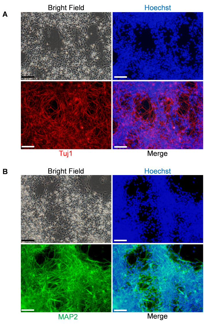
Human induced pluripotent stem cells (iPSCs) and their progeny displaying tissue-specific characteristics have paved the way for regenerative medicine and research in various fields such as the elucidation of the pathological mechanism of diseases and the discovery of drug candidates. iPSC-derived neurons are particularly valuable as it is difficult to analyze neural cells obtained from the central nervous system in humans. For neuronal induction with iPSCs, one of the commonly used approaches is the isolation and expansion of neural rosettes, following the formation of embryonic bodies (EBs). However, this process is laborious, inefficient, and requires further purification of the cells. To overcome these limitations, we have developed an efficient neural induction method that allows for the generation of neural stem/progenitor cells (NSCs/NPCs) from iPSCs within 7 days and of functional mature neurons. Our method yields a PAX6-positive homogeneous cell population, a cortical NSCs/NPCs, and the resultant NSCs/NPCs can be cryopreserved, expanded, and differentiated into functional mature neurons. Moreover, our protocol will be less expensive than other methods since the protocol requires fewer neural supplements during neural induction. This article also presents the FM1-43 imaging assay, which is useful for the presynaptic assessment of the iPSCs-derived human neurons. This protocol provides a quick and simplified way to generate NSCs/NPCs and neurons, enabling researchers to establish in vitro cellular models to study brain disease pathology.
Keywords: FM1-43; Human Induced Pluripotent Stem Cell; Neural Induction; Neural Progenitor Cell; Neural Stem Cell; Neuron.
Copyright © 2021 The Authors; exclusive licensee Bio-protocol LLC.
Conflict of interest statement
Competing interestsThe authors declare that they have no conflict of interest.
Figures


References
-
- Dimos J. T., Rodolfa K. T., Niakan K. K., Weisenthal L. M., Mitsumoto H., Chung W., Croft G. F., Saphier G., Leibel R., Goland R., Wichterle H., Henderson C. E. and Eggan K.(2008). Induced pluripotent stem cells generated from patients with ALS can be differentiated into motor neurons. Science 321(5893): 1218-1221. - PubMed
-
- Dunn N. R., Vincent S. D., Oxburgh L., Robertson E. J. and Bikoff E. K.(2004). Combinatorial activities of Smad2 and Smad3 regulate mesoderm formation and patterning in the mouse embryo. Development 131(8): 1717-1728. - PubMed
-
- Era T., Izumi N., Hayashi M., Tada S., Nishikawa S. and Nishikawa S. I.(2008). Multiple mesoderm subsets give rise to endothelial cells, whereas hematopoietic cells are differentiated only from a restricted subset in embryonic stem cell differentiation culture. Stem Cells 26(2): 401-411. - PubMed
-
- Falk A., Koch P., Kesavan J., Takashima Y., Ladewig J., Alexander M., Wiskow O., Tailor J., Trotter M., Pollard S., Smith A. and Brustle O.(2012). Capture of neuroepithelial-like stem cells from pluripotent stem cells provides a versatile system for in vitro production of human neurons . PLoS One 7(1): e29597. - PMC - PubMed
LinkOut - more resources
Full Text Sources
Other Literature Sources
