

Use of porcine acellular dermal matrix to repair lung Hernia after minithoracotomy: A case report with 6-Year follow-up
- PMID: 33732854
- PMCID: PMC7937533
- DOI: 10.1016/j.jpra.2021.01.012
Use of porcine acellular dermal matrix to repair lung Hernia after minithoracotomy: A case report with 6-Year follow-up
Abstract
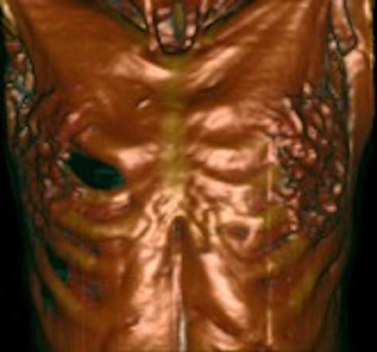
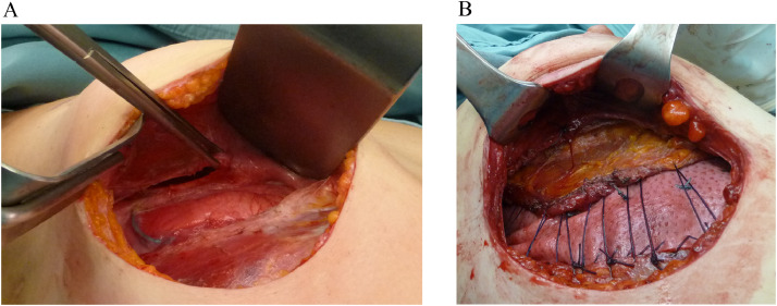
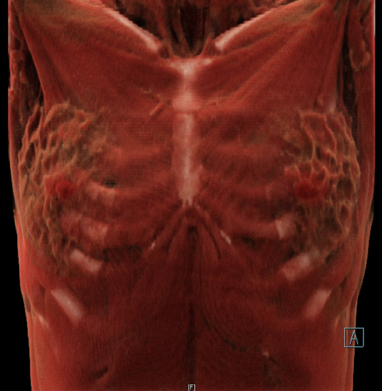
Lung hernia following minimally invasive cardiac surgery is rare with few reported cases in the literature. Surgical repair is debated, and several methods have been described including a variety of synthetic and biological materials. We report a case of a 36-year-old woman who developed lung hernia and a strong retraction of the pectoralis major muscle after minithoracotomy that was performed for mitral valve surgery. The herniated lung was reduced and the chest wall defect was repaired with a non-cross linked acellular dermal matrix (ADM) anchored to the thoracic wall. At a 6-year follow-up, she was asymptomatic and without recurrence of the hernia. Our experience suggests that ADMs are a safe and reliable surgical technique for lung hernia repair due to their biological and mechanical properties, even in those secondary hernias to minithoracotomy where a complete muscle coverage of the matrix could not be provided.
Keywords: Acellular dermal matrix; Chest wall reconstruction; Lung hernia; Minimally invasive valve surgery; Minithoracotomy; Strattice.
© 2021 The Authors. Published by Elsevier Ltd on behalf of British Association of Plastic, Reconstructive and Aesthetic Surgeons.
Conflict of interest statement
The authors declare no conflicts of interest.
Figures
Similar articles
-
Porcine incisional hernia model: Evaluation of biologically derived intact extracellular matrix repairs.J Tissue Eng. 2013 Oct 10;4:2041731413508771. doi: 10.1177/2041731413508771. eCollection 2013. J Tissue Eng. 2013. PMID: 24555008 Free PMC article.
-
Acellular Dermal Matrix Susceptibility to Collagen Digestion: Effect on Mechanics and Host Response.Tissue Eng Part A. 2023 May;29(9-10):269-281. doi: 10.1089/ten.TEA.2022.0155. Epub 2023 Mar 16. Tissue Eng Part A. 2023. PMID: 36641639
-
Pulmonary hernia secondary to limited access for mitral valve surgery and repaired by video thoracoscopic surgery.Interact Cardiovasc Thorac Surg. 2009 Jan;8(1):111-3. doi: 10.1510/icvts.2008.190744. Epub 2008 Oct 23. Interact Cardiovasc Thorac Surg. 2009. PMID: 18948304
-
Complex Ventral Hernia Repair with Acellular Dermal Matrices: Clinical and Quality of Life Outcomes.Am Surg. 2017 Feb 1;83(2):141-147. Am Surg. 2017. PMID: 28228200
-
Open and Laparo-Endoscopic Repair of Incarcerated Abdominal Wall Hernias by the Use of Biological and Biosynthetic Meshes.Front Surg. 2016 Feb 25;3:10. doi: 10.3389/fsurg.2016.00010. eCollection 2016. Front Surg. 2016. PMID: 26942182 Free PMC article. Review.
Cited by
-
Acellular dermal matrix in reconstructive surgery: Applications, benefits, and cost.Front Transplant. 2023 Mar 10;2:1133806. doi: 10.3389/frtra.2023.1133806. eCollection 2023. Front Transplant. 2023. PMID: 38993878 Free PMC article. Review.
-
Human Acellular Dermal Matrix in Reconstructive Surgery-A Review.Biomedicines. 2022 Nov 9;10(11):2870. doi: 10.3390/biomedicines10112870. Biomedicines. 2022. PMID: 36359387 Free PMC article. Review.
References
Publication types
LinkOut - more resources
Full Text Sources
Other Literature Sources
