. 2021 Jan 25;3(3):123-135.
doi: 10.1096/fba.2020-00039. eCollection 2021 Mar.

Beclin-1 improves mitochondria-associated membranes in the heart during endotoxemia

Affiliations

Beclin-1 improves mitochondria-associated membranes in the heart during endotoxemia

Yuxiao Sun et al. FASEB Bioadv. .

Abstract

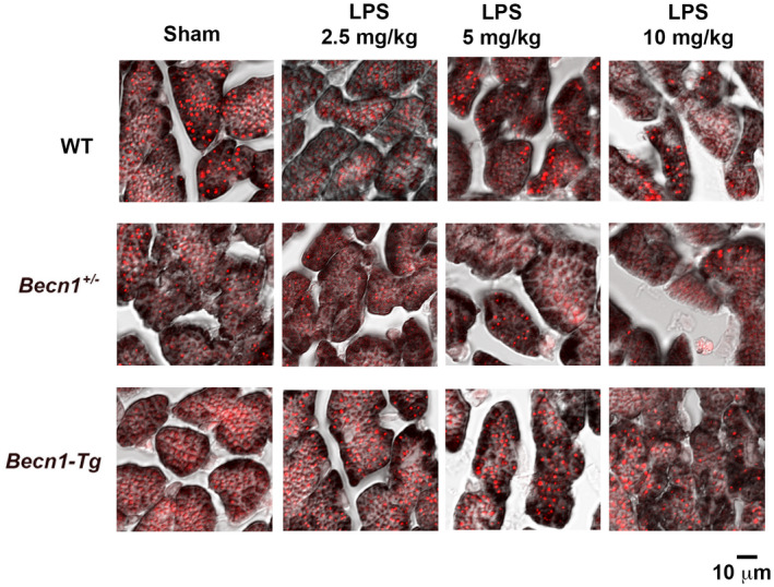
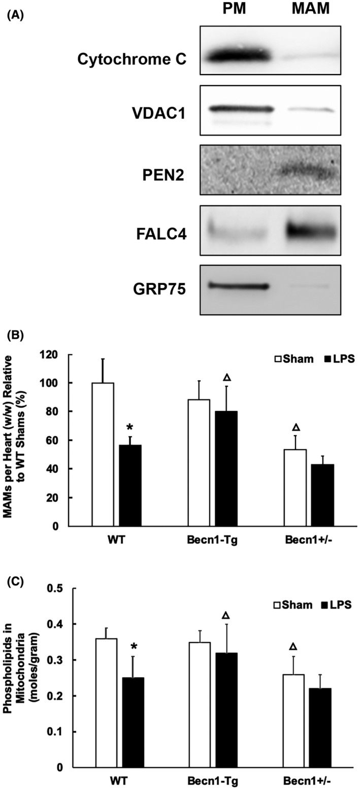
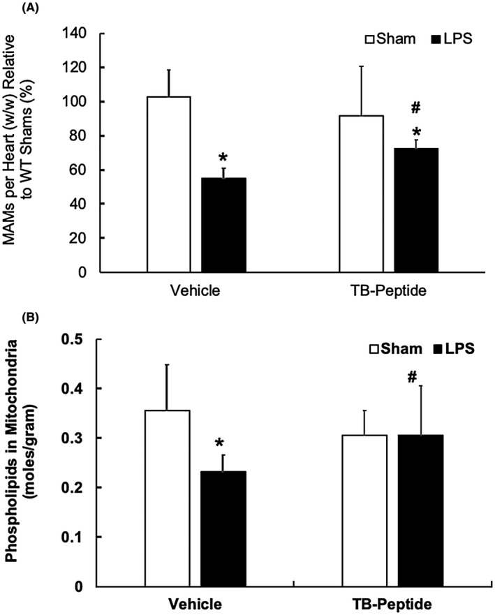
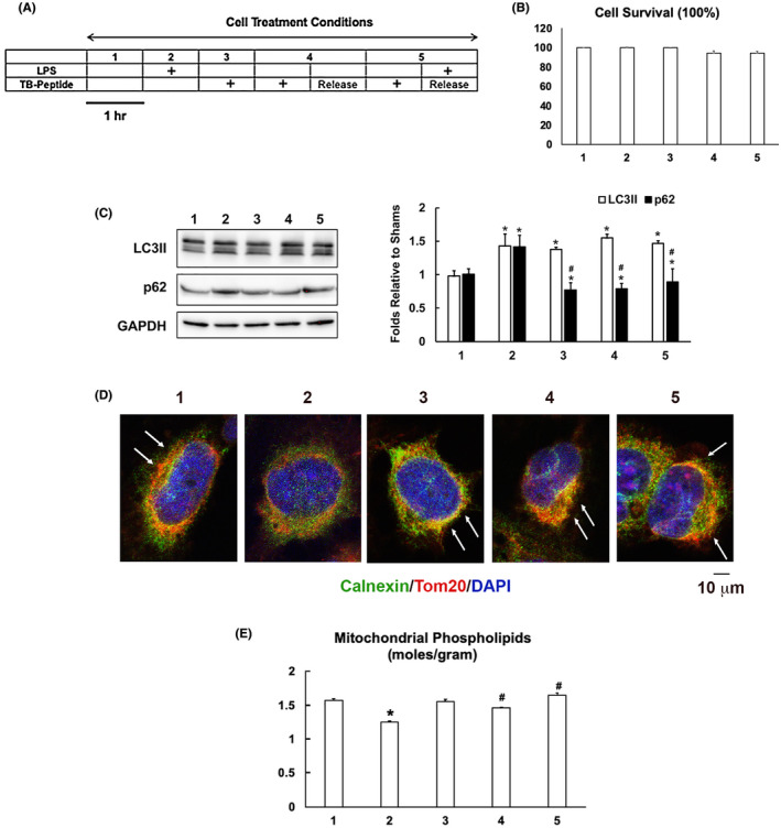
Mitochondria-associated membranes (MAMs) are essential to mitochondria. This study was to determine whether endotoxemia rearranges MAMs in the heart, and whether Beclin-1 regulates this process. Wild-type mice and mice with a cardiac-specific overexpression of Beclin-1 (Becn1-Tg), or a heterozygous knockout of Beclin-1 (Becn1 +/-) were given lipopolysaccharide (LPS) challenge. In the heart, the ultrastructure of MAMs was examined by electron microscopy and the histology evaluated by immunostaining. Additionally, MAMs were isolated by ultracentrifugation, and their content and function were quantified. The effects of Beclin-1-activating peptide (TB-peptide) on MAMs were also examined. Data showed that endotoxemia decreased both the total mass and the function of MAMs, and these deficiencies became worse in Becn1 +/- mice but were alleviated in Becn1-Tg and TB-peptide-treated mice. Responses of myocardial MAMs to LPS and to TB-peptide were additionally examined in AC16 human cardiomyocytes. In vitro findings recaptured the effects of LPS and TB-peptide in cardiomyocytes; the challenge of LPS reduced the level and activity of MAMs, and TB-peptide attenuated this defect. Together, the results suggest a new function of Beclin-1 in improving cardiac MAMs during endotoxemia, providing a mechanism for the previously identified role of Beclin-1 in protection of mitochondria and cardiac function.

Keywords: LPS; MAMs; beclin‐1; cardiac dysfunction; sepsis.


Figures

FIGURE 1
FIGURE 1
LPS‐induced changes in the structure of cardiac MAMs in WT, Becn1‐Tg, and Becn1 +/− mice. Mice were given indicated doses of LPS via i.p., and heart tissues were harvested 18 hours later. A, Ultrastructure of mitochondria and MAMs was observed by transmission electron microscope. Red arrows indicate the structure of MAMs. B, Heart tissue sections were co‐immune‐stained with the ER marker calreticulin (red) and mitochondria marker VDAC1 (green). Overlay color in yellow indicates the levels of MAMs. All images are representative of n ≥ 3 animals per group
FIGURE 2
FIGURE 2
LPS‐induced changes in mitochondria and lipid contents in WT, Becn1‐Tg, and Becn1 +/− mice. Mice were given indicated doses of LPS via i.p., and heart tissues were harvested 18 hours later. Heart tissue sections were co‐stained with mitochondria marker VDAC1 (brown) and lipid marker Nile red (red). All images are representative of n ≥ 3 animals per group
FIGURE 3
FIGURE 3
LPS‐induced changes in the mass and function of cardiac MAMs in WT, Becn1‐Tg, and Becn1 +/− mice. Mice were given 5 mg/kg LPS via i.p., and heart tissues were harvested 18 hours later. Fractions of pure mitochondria (PM) and MAMs were prepared by ultracentrifugation from the heart tissue. A, Successful isolation of MAMs and PM from the hearts of WT mice was demonstrated by Western blots detecting marker proteins of mitochondria and ER. B, The amount of each fraction of MAMs were measured and results were normalized by tissue weight. C, Levels of phospholipids in mitochondrial fractions were quantified and results were normalized by the amount of protein. In B and C, values are means ±SEM. Significant differences are shown as * for sham vs LPS‐treated and Δ for WT vs Becn1‐Tg or Becn1 +/− groups (P < .05, n = 4‐6, unpaired t test)
FIGURE 4
FIGURE 4
Effects of TB‐peptide on cardiac MAMs in LPS‐challenged mice. WT mice were given 5 mg/kg LPS i.p., then TB‐peptide, 16 mg/kg, was administered i.p. 30 minutes post LPS challenge. Heart tissues were collected 18 h post LPS challenge, and fractions of mitochondria and MAMs were prepared by ultracentrifugation. A, The amount of each fraction of MAMs was measured and results were normalized by tissue weight. B, Levels of phospholipids in mitochondrial fractions were quantified and results were normalized by the amount of protein. All values are means ±SEM. Significant differences are shown as * for sham vs LPS‐treated and # for without vs with TB‐peptide (P < .05, n = 4‐6, unpaired t test)
FIGURE 5
FIGURE 5
TB‐peptide promotes autophagy and protects MAMs in human cardiomyocyte challenged by LPS. A, AC16 cells were cultured until 80%‐90% confluency and treated with the conditions as illustrated. B, The percentage of cell survival was calculated based on the analysis of cytotoxicity. C, Levels of LC3II and p62 in cell lysates were examined by western blot and quantified by densitometry. GAPDH was used as a loading control. D, Cells were co‐immune‐stained with the ER marker calnexin (green) and mitochondria marker Tom 20 (red). Staining of nucleus by DAPI is shown in blue. Overlay areas of mitochondria–ER contact shown in the color yellow indicates the levels of MAMs and are labeled with white arrows. All images are representative of ≥3 independent experiments. E, Levels of phospholipids in mitochondrial fractions were quantified and results were normalized by the amount of protein. All values are means ±SEM. Significant differences are shown as * for sham vs LPS‐treated and # for without vs with TB‐peptide (p < .05, n = 3‐5, unpaired t test)

Similar articles

Cited by

References

    1. Singer M, Deutschman CS, Seymour CW, et al. The Third International Consensus Definitions for Sepsis and Septic Shock (Sepsis‐3). J Am Med Assoc. 2016;315:801–810. - PMC - PubMed
    1. Levy MM, Dellinger RP, Townsend SR, et al. The Surviving Sepsis Campaign: results of an international guideline‐based performance improvement program targeting severe sepsis*. Critical Care Med. 2010;38(2):367–374. - PubMed
    1. Iwashyna TJ, Ely EW, Smith DM, Langa KM. Long‐term cognitive impairment and functional disability among survivors of severe sepsis. J Am Med Assoc. 2010;304:1787–1794. - PMC - PubMed
    1. Walley KR. Sepsis‐induced myocardial dysfunction. Curr Opin Crit Care. 2018;24:292–299. - PubMed
    1. Zanotti‐Cavazzoni SL, Hollenberg SM. Cardiac dysfunction in severe sepsis and septic shock. Curr Opin Crit Care. 2009;15:392–397. - PubMed

LinkOut - more resources