. 2021 Feb 17;24(3):102201.
doi: 10.1016/j.isci.2021.102201. eCollection 2021 Mar 19.

Structure of a type IV CRISPR-Cas ribonucleoprotein complex

Affiliations

Structure of a type IV CRISPR-Cas ribonucleoprotein complex

Yi Zhou et al. iScience. .

Abstract

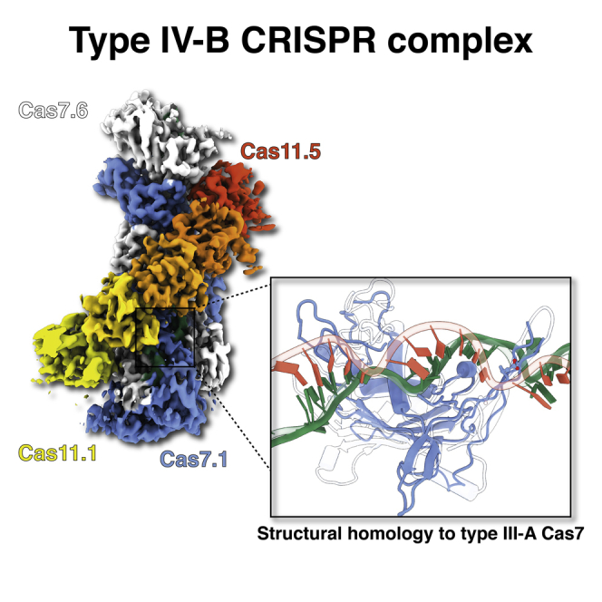
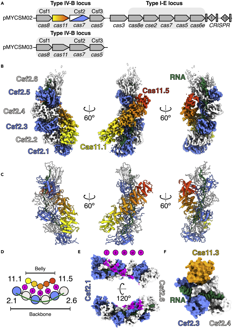
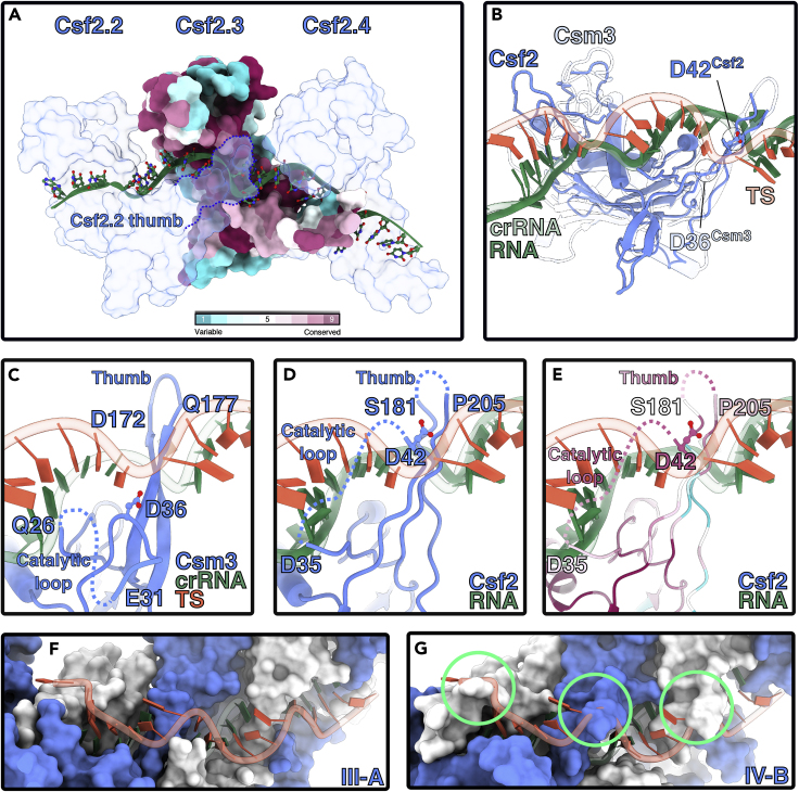
We reveal the cryo-electron microscopy structure of a type IV-B CRISPR ribonucleoprotein (RNP) complex (Csf) at 3.9-Å resolution. The complex best resembles the type III-A CRISPR Csm effector complex, consisting of a Cas7-like (Csf2) filament intertwined with a small subunit (Cas11) filament, but the complex lacks subunits for RNA processing and target DNA cleavage. Surprisingly, instead of assembling around a CRISPR-derived RNA (crRNA), the complex assembles upon heterogeneous RNA of a regular length arranged in a pseudo-A-form configuration. These findings provide a high-resolution glimpse into the assembly and function of enigmatic type IV CRISPR systems, expanding our understanding of class I CRISPR-Cas system architecture, and suggesting a function for type IV-B RNPs that may be distinct from other class 1 CRISPR-associated systems.

Keywords: Biological Sciences; Structural Biology.


Conflict of interest statement

The authors declare no competing interests.

Figures

None
Graphical abstract
Figure 1
Figure 1
Structure of type IV-B CRISPR complex See also Figures S1–S4 and Table S1. (A) M. sp. JS623 plasmid-encoded CRISPR operons. Top: Type IV-B and I-E CRISPR loci present on pMCYCM02 megaplasmid. Bottom: Additional type IV-B locus encoded by pMCYCM03 megaplasmid. Genes predicted to encode RNP complex subunits are indicated with a gray rectangle. (B) 3.9 Å-resolution cryo-EM reconstruction of type IV-B CRISPR complex. Cas7 subunits are colored blue and white, and five Cas11 subunits are colored as a yellow-orange-red gradient. Csf-bound RNA is green. (C) Refined model for the Csf effector complex derived from the cryo-EM maps shown in (B). (D) Schematic of Cas7-Cas11 interactions. Five Csf2-Cas11 interactions occur in this complex (labeled i – v). (E) Positions of Cas11 contacts on Csf2 backbone, colored magenta as shown in panel D. Cas11 sits upon the Csf2-Csf2 interface. (F) Cas11 binds at the interface with buried surface area of 505 Å2 (150 Å2 and 355 Å2 with Csf2.3 and Csf2.4, respectively). Cas11 is completely occluded from bound RNA. Csf2 subunits are intimately connected (1021 Å2) and make a network of contacts with bound RNA (∼1200 Å2 buried surface area per Csf2 subunit).
Figure 2
Figure 2
RNA-binding by type IV-B Cas7 See also Figures S5–S7. (A) RNA (green) binding site runs across the palms of Csf2 subunits. Csf2.3 is colored according to conservation. The “thumb” of the n-1 Csf2 (i.e. Csf2.2) protrudes into the backbone of bound RNA (solid green), inducing a kink. (B) Alignment of type III-A backbone subunit Csm3 (PDB 607i, transparent) with Csf2 (solid blue). Csm3 and Csf2 align with an r.m.s.d. of 2.9Å, with a Dali server Dali server Z score 14.1. Csf2-bound RNA binds in the same conformation as crRNA (transparent green) to Csm3 (RMSD of 1.5 Å). Catalytic residue Asp36Csm3 and putative catalytic residue Asp42Csf2 side chains are located near the target strand (TS - transparent red), bound to the type III crRNA (transparent green). (C) Residues flanking unstructured catalytic loop (27–35) and apical loop of Csm3 thumb also interact with the TS. Catalytic residue D36 is shown for clarity. (D) Putative interactions with Csf2 and TS, based on alignments with the Csm complex. (E) Putative interactions colored by conservation. The Csf2 thumb contains a flexible 20 residue insertion, not visible in our cryo-EM map. (F), Path of TS bound by type III-A Csm complex. (G) Putative path of TS along IV-B. Severe classes with TS and Csf2 are circled in green.

References

    1. Chowdhury S., Carter J., Rollins M.C.F., Golden S.M., Jackson R.N., Hoffmann C., Nosaka L., Bondy-Denomy J., Maxwell K.L., Davidson A.R. Structure reveals mechanisms of viral suppressors that intercept a CRISPR RNA-guided surveillance complex. Cell. 2017;169:47–57.e11. - PMC - PubMed
    1. Crowley V.M., Catching A., Taylor H.N., Borges A.L., Metcalf J., Bondy-Denomy J., Jackson R.N. A type IV-A CRISPR-cas system in Pseudomonas aeruginosa mediates RNA-guided plasmid interference in vivo. Cris. J. 2019;2:434–440. - PMC - PubMed
    1. Faure G., Shmakov S.A., Yan W.X., Cheng D.R., Scott D.A., Peters J.E., Makarova K.S., Koonin E.V. CRISPR–Cas in mobile genetic elements: counter-defence and beyond. Nat. Rev. Microbiol. 2019;17:513–525. - PMC - PubMed
    1. Halpin-Healy T.S., Klompe S.E., Sternberg S.H., Fernández I.S. Structural basis of DNA targeting by a transposon-encoded CRISPR–Cas system. Nature. 2020;577:271–274. - PubMed
    1. Hille F., Richter H., Wong S.P., Bratovič M., Ressel S., Charpentier E. The biology of CRISPR-cas: backward and forward. Cell. 2018;172:1239–1259. - PubMed

LinkOut - more resources