. 2021 Feb 23:7:100089.
doi: 10.1016/j.jvacx.2021.100089. eCollection 2021 Apr.

The Leptospiral General Secretory Protein D (GspD), a secretin, elicits complement-independent bactericidal antibody against diverse Leptospira species and serovars

Affiliations

The Leptospiral General Secretory Protein D (GspD), a secretin, elicits complement-independent bactericidal antibody against diverse Leptospira species and serovars

Eja Schuler et al. Vaccine X. .

Abstract

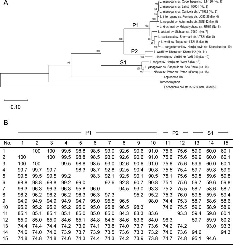
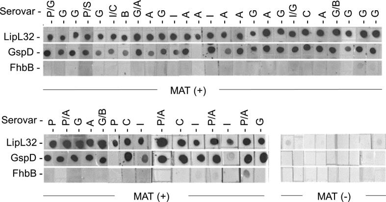
Leptospirosis, the most common zoonotic infection worldwide, is a multi-system disorder affecting the kidney, liver, and lungs. Infections can be asymptomatic, self-limiting or progress to multi-organ system failure and pulmonary hemorrhage. The incidence of canine and human leptospirosis is steadily increasing worldwide. At least sixty-four Leptospira species and several hundred lipopolysaccharide-based serovars have been defined. Preventive vaccines are available for use in veterinary medicine and limited use in humans in some countries. All commercially available vaccines are bacterin formulations that consist of a combination of laboratory cultivated strains of different lipopolysaccharide serotypes. The development of a broadly protective subunit vaccine would represent a significant step forward in efforts to combat leptospirosis in humans, livestock, and companion animals worldwide. Here we investigate the potential of General secretory protein D (GspD; LIC11570), a secretin, to serve as a possible antigen in a multi-valent vaccine formulation. GspD is conserved, expressed in vitro, antigenic during infection and elicits antibody with complement independent bactericidal activity. Importantly, antibody to GspD is bactericidal against diverse Leptospira species of the P1 subclade. Epitope mapping localized the bactericidal epitopes to the N-terminal N0 domain of GspD. The data within support further exploration of GspD as a candidate for inclusion in a next generation multi-protein subunit vaccine.

Keywords: Canine; GspD; Leptospirosis; Secretin; Spirochetes; Type 2 secretion.


Conflict of interest statement

The authors declare that they have no known competing financial interests or personal relationships that could have appeared to influence the work reported in this paper.

Figures

Fig. 1
Fig. 1
Phylogenetic analyses and pairwise sequence comparisons of GspD amino acid sequences. Panel A presents a phylogenetic tree constructed using GspD amino acid sequences as detailed in the text. Representative GspD sequences from Leptospira subclades P1, P2, and S1 (as indicated) were included. Leptonema illini, Turneriella parva and Escherichia coli str. K-12 served as outgroups. The results of bootstrap analyses are indicated on each node and the scale bar is shown in the lower left. Note that the numbers (No.) in parentheses that follow each isolate designation correspond to those used in the distance matrix table (Panel B). Panel B presents the GspD percent amino acid similarity and identity values (lower and upper quadrants, respectively). The subclade classification of each isolate of origin is indicated along the top of panel B. All accession numbers can be found in Table 1.
Fig. 2
Fig. 2
In vitro expression and cellular localization of GspD. Panel A presents an immunoblot in which whole cell lysates of diverse Leptospira isolates were screened with anti-GspD antiserum as detailed in the text. In panel B, L. interrogans serovar Copenhageni strain Fiocruz L1-130 was subjected to Triton X-114 extraction and phase partitioning. The resulting fractions were separated by SDS-PAGE, transferred to PVDF membranes and screened with antiserum (1:1000 dilution) as indicated below each immunoblot. The left panel of Panel B presents a Coomassie Brilliant Blue stained SDS-PAGE gel. Antisera against LipL32 and Qlp42 served as partitioning controls for outer membrane and periplasmic proteins, respectively. Abbreviations are as follows: whole cell lysate (WCL), protoplasmic cylinder (PC), aqueous phase (AQ), and detergent phase (DET). Molecular weight standards are indicated to the left. All methods were as detailed in the text.
Fig. 3
Fig. 3
GspD is antigenic during natural infection in client-owned canines. Dot-blots of full length recombinant proteins (indicated to the left) were screened with sera from MAT positive and MAT negative client owned dogs (1:200 dilution) as indicated below each panel. For each serum sample, the serovar(s) that yielded the the highest MAT titers are indicated above each dot-blot. Abbreviations were as follows: Pomona (P), Icterohaemorrhagiae (I), Canicola (C), Grippotyphosa (G), Hardjo-bovis (H), Bratislava (B), Autumnalis (A), and Sejroe (S). For sera that had equal titers to two serovars, both serovar abbreviations are provided. LipL32 was included as a positive control and the T. denticola FhbB protein served as a negative control. Molecular weight standards are indicated on the left.
Fig. 4
Fig. 4
Anti-GspD antisera is bactericidal and kills diverse strains through a complement independent mechanism. The bactericidal activity of rat anti-GspD antiserum against diverse Leptospira strains (as indicated in the figure key) was assessed as described in the methods. Abbreviations are as follows: NHS (complement certified normal human serum), HI-NHS (heat inactivated normal human sera), and Rat PI (rat pre-immune serum). Equivalent numbers of cells were incubated with NHS, HI-NHS, rat PI, or anti-GspD antisera as indicated below the figure. Percent killing was determined by visual examination of cell numbers per field of view using dark field microscopy as detailed in the text. Differences in cell counts per field of view in the negative control (NHS + rat PI) versus in the treatment groups (NHS + Anti-GspD and HI-NHS + Anti-GspD) were assessed using the two-tailed student’s t-test (* indicates P ≤ .05).
Fig. 5
Fig. 5
Localization of the bactericidal epitopes of GspD to the N-terminal domain. In panel A, full-length (FL) GspD and sub-fragments (indicated as F1 through F5) were screened with serum from nine individual MAT + canines (1:100 dilution) by ELISA. IgG levels to each protein were determined as detailed in the text. The data were assessed using the two-tailed student’s t test based on the mean value for each group (* indicates P < .05). In panel B, anti-GspD antisera was adsorbed with full length recombinant GspD FL or the F1 through F5 fragments (as indicated along the x axis). The adsorbed sera were then used in bactericidal assays in the presence of complement preserved normal human serum (NHS) and percent killing was determined as described in the text. Cells incubated with NHS and rat pre-immune (PI) serum alone served as a negative control. Cells incubated with NHS and non-adsorbed anti-GspD antiserum (GspD antisera) served as a positive control. Significance was evaluated by comparing cells per field of view of each treatment group with the GspD antisera alone control sample using the two-tailed student’s t test (* indicates P < .05).

Similar articles

Cited by

References

    1. Divers T.J., Chang Y.F., Irby N.L., Smith J.L., Carter C.N. Leptospirosis: An important infectious disease in North American horses. Equine Vet J. 2019;51:287–292. - PubMed
    1. Bulach D., Adler B. Leptospiral Genomics and Pathogenesis. Curr Top Microbiol Immunol. 2018;415:189–214. - PubMed
    1. Vincent A.T., Schiettekatte O., Goarant C., Neela V.K., Bernet E., Thibeaux R. Revisiting the taxonomy and evolution of pathogenicity of the genus Leptospira through the prism of genomics. PLoS Negl Trop Dis. 2019;13 - PMC - PubMed
    1. Adler B., de la Pena Moctezuma A. Leptospira and leptospirosis. Vet Microbiol. 2010;140:287–296. - PubMed
    1. Cilia G., Bertelloni F., Fratini F. Leptospira Infections in Domestic and Wild Animals. Pathogens. 2020;9 - PMC - PubMed

LinkOut - more resources