. 2021 Apr;78(7):3709-3724.
doi: 10.1007/s00018-021-03795-w. Epub 2021 Mar 17.

RNA and DNA G-quadruplexes bind to human dicer and inhibit its activity

Affiliations

RNA and DNA G-quadruplexes bind to human dicer and inhibit its activity

Natalia Koralewska et al. Cell Mol Life Sci. 2021 Apr.

Abstract

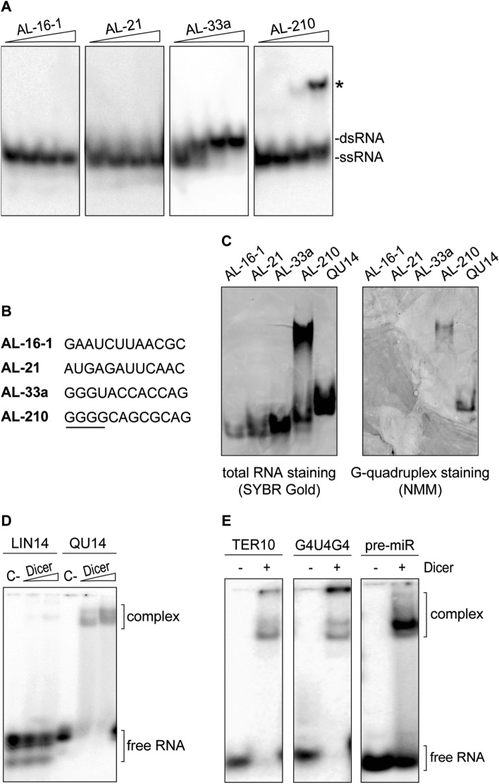
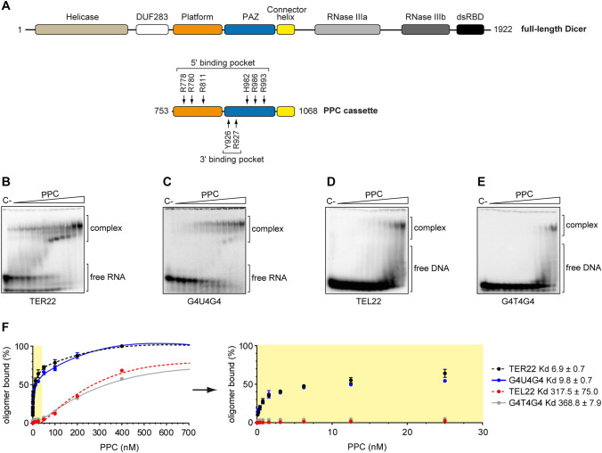
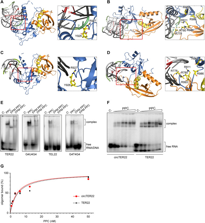
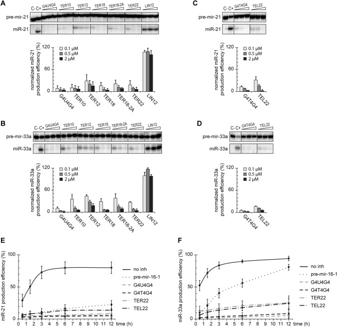
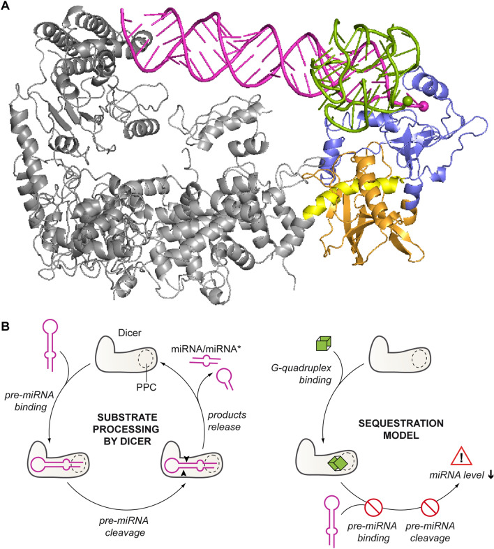
Guanine (G)-rich single-stranded nucleic acids can adopt G-quadruplex structures. Accumulating evidence indicates that G-quadruplexes serve important regulatory roles in fundamental biological processes such as DNA replication, transcription, and translation, while aberrant G-quadruplex formation is linked to genome instability and cancer. Understanding the biological functions played by G-quadruplexes requires detailed knowledge of their protein interactome. Here, we report that both RNA and DNA G-quadruplexes are bound by human Dicer in vitro. Using in vitro binding assays, mutation studies, and computational modeling we demonstrate that G-quadruplexes can interact with the Platform-PAZ-Connector helix cassette of Dicer, the region responsible for anchoring microRNA precursors (pre-miRNAs). Consequently, we show that G-quadruplexes efficiently and stably inhibit the cleavage of pre-miRNA by Dicer. Our data highlight the potential of human Dicer for binding of G-quadruplexes and allow us to propose a G-quadruplex-driven sequestration mechanism of Dicer regulation.

Keywords: Dicer PPC cassette; Dicer inhibition; MiRNA biogenesis; PAZ domain; Regulation of enzyme activity; Ribonucleoprotein complexes.


Conflict of interest statement

The authors declare no conflict of interest.

Figures

Fig. 1
Fig. 1
hDicer binds RNA G-quadruplexes. a Native PAGE analysis of the mixtures of unlabeled and 5′-32P-labeled RNA 12-mers (AL). Triangles represent increasing amounts of a given oligomer (0.01, 0.1, 1, 10 µM). Bands corresponding to single- and double-stranded forms of the oligomers are indicated. b Comparison of the sequences of RNA 12-mers used in the study. A guanine tract in the sequence of AL-210 is underlined. c Native PAGE analysis of RNA 12-mers (AL) and a control 14-nt G-quadruplex (QU14) (100 pmol each). Gels were treated with nucleic acid stain SYBR Gold (left) or G-quadruplex-specific dye, NMM (right). d EMSAs with hDicer and the 5′-32P-labeled 14-nt RNA not adopting a G-quadruplex structure (LIN14), or the 5′-32P-labeled RNA G-quadruplex (QU14). C− a control sample with no protein. Triangles represent increasing amounts of hDicer (100, 250 nM). e EMSAs with hDicer (250 nM) and the 5′-32P-labeled RNA G-quadruplex (TER10 or G4U4G4) or a control pre-miRNA. ± refers to Dicer presence in the reaction mixture
Fig. 2
Fig. 2
The PPC cassette of hDicer binds RNA and DNA G-quadruplexes. a Domain architecture of hDicer (top) and PPC cassette (below). Arrows indicate the RNA-interacting residues within the 5′- and 3′-pocket. b, c EMSAs with PPC and the 5′-32P-labeled RNA G-quadruplexes TER22 b and G4U4G4 c. C− a control sample with no protein. Triangles represent increasing amounts of PPC (0.1, 0.2, 0.4, 0.8, 1.6, 3.125, 6.25, 12.5, 25, 50, 100, 200, 400 nM). The Kd of RNA–PPC complexes was determined from the binding isotherms by curve-fitting using nonlinear regression. Error bars represent SD from three separate experiments. d, e EMSAs with PPC and the 5′-32P-labeled DNA G-quadruplexes TEL22 d and G4T4G4 e. C− a control sample with no protein. Triangles represent increasing amounts of PPC (0.1, 0.2, 0.4, 0.8, 1.6, 3.125, 6.25, 12.5, 25, 50, 100, 200, 400 nM). The Kd values of DNA–PPC complexes were determined from the binding isotherms by curve fitting using nonlinear regression. Error bars represent SD from three separate experiments. f Quantitative analysis of the binding assay between PPC and 5′-32P-labeled RNA/DNA G-quadruplexes. Error bars represent SD from three separate experiments
Fig. 3
Fig. 3
Both the 3′- and 5′-pocket of hDicer PPC cassette are involved in the binding of G-quadruplexes. a 3D model of TER10 forming a dimer of bimolecular G-quadruplexes bound to the 3′-pocket of hDicer PPC cassette. The 3′- and 5′-ends of the oligonucleotide are colored in green and red, respectively. The Platform is colored in orange, PAZ in blue and Connector helix in yellow. The same color coding is maintained in subpanels bd of this b 3D model of TER10 forming a dimer of bimolecular G-quadruplexes bound to the 5′-pocket of hDicer PPC cassette. c 3D model of G4T4G4 forming a bimolecular G-quadruplex bound to the 3′-pocket of hDicer PPC cassette. d 3D model of G4T4G4 forming a bimolecular G-quadruplex bound to the 5′-pocket of hDicer PPC cassette. e EMSAs with PPC, PPC 5′- and 3′-pocket mutants (5′PM, 3′PM), and 5′-32P-labeled G-quadruplexes (TER22, G4U4G4, TEL22, G4T4G4). 500 nM protein and 10,000 cpm (approximately 5 nM) of RNA/DNA were used per lane. C− a control sample with no protein. f EMSAs of binding between PPC and TER22 with free ends (TER22) or TER22 with ligated ends (circTER22). Triangles represent the increasing amount of PPC (0.8, 1.6, 3.125, 6.25, 12.5, 50 nM). C− a control sample with no protein. g Quantitative analysis of the binding assay between PPC and TER22/circTER22 G-quadruplexes. Error bars represent SD from three separate experiments
Fig. 4
Fig. 4
RNA G-quadruplexes bound by the PPC cassette retain their structure. a EMSAs with 500 nM PPC and TER22 or LIN32, which does not adopt a G-quadruplex structure. Both RNA oligonucleotides were labeled at the 5′-end with a G-quadruplex specific probe (o-BMVC). ± indicates the presence or absence of PPC, or the RNA oligomer. Triangles represent increasing amounts of the RNA oligomer (7.5, 15, 22.5, 30 µM). To visualize RNA adopting G-quadruplex structures, the gel was exposed to 532 nm light. b The same gel as presented in panel a exposed to 473 nm light after staining with SYBR Gold to visualize total RNA
Fig. 5
Fig. 5
G-quadruplexes inhibit cleavage of pre-miRNA by hDicer in a dose-dependent manner. a, b Inhibition assay to assess the effect of RNA G-quadruplexes on the cleavage of pre-mir-21 (a) or pre-mir-33a (b) by hDicer. Reactions were carried out for 30 min at 37 °C under the low-turnover conditions. LIN12—a control 12-mer not adopting a G-quadruplex structure, C− a sample with no protein, nor inhibitor added, C+ a sample with hDicer, without inhibitor. Graphs show miRNA production efficiency normalized to the level of miRNA generated in C+. Error bars represent SD from three separate experiments. See also Figure S4B, C for full gel images. The reproducible results were obtained using at least two batches of recombinant hDicer. c, d The results of the inhibition assay performed similarly as in (a, b) for selected DNA G-quadruplexes and pre-mir-21 (c) or pre-mir-33a (d). See also Figure S4D, E for full gel images. The reproducible results were obtained using at least two batches of recombinant hDicer. e, f Quantitative analysis of the time course of hDicer inhibition by G-quadruplexes in reactions with pre-mir-21 (e) or pre-mir-33a (f). Error bars represent SD from three separate experiments; see also Figure S5A–D for representative gel images. The reproducible results were obtained using at least two batches of recombinant hDicer
Fig. 6
Fig. 6
Both pre-miRNA and G-quadruplexes are anchored within the same region of hDicer PPC cassette. a Superposed structures of hDicer in complex with a pre-miRNA substrate (PDB entry 5ZAL, does not include 15 nt from the apical loop of the pre-miRNA) and PPC cassette in complex with TER10. The Platform is colored in orange, PAZ in blue and Connector helix in yellow, the remaining portion of hDicer is presented in grey; pre-miRNA—in magenta, and TER10—in green. 3′ ends of pre-miRNA and TER10 within the 3′-binding pocket of PAZ are marked as spheres. b Putative mechanism of sequestration-dependent regulation of Dicer activity by G-quadruplexes. Dicer anchors pre-miRNA ends within the PPC binding pockets and cleaves precursors to release miRNA products (left). G-quadruplexes compete with pre-miRNA for binding to Dicer. The enzyme sequestered in complex with a G-quadruplex cannot bind pre-miRNA and does not generate miRNA (right)

Similar articles

Cited by

References

    1. Court DL, Gan J, Liang YH, Shaw GX, Tropea JE, Costantino N, Waugh DS, Ji X. RNase III: genetics and function; structure and mechanism. Annu Rev Genet. 2013;47:405–431. doi: 10.1146/annurev-genet-110711-155618. - DOI - PMC - PubMed
    1. Lau PW, Guiley KZ, De N, Potter CS, Carragher B, MacRae IJ. The molecular architecture of human Dicer. Nat Struct Mol Biol. 2012;19(4):436–440. doi: 10.1038/nsmb.2268. - DOI - PMC - PubMed
    1. Tian Y, Simanshu DK, Ma JB, Park JE, Heo I, Kim VN, Patel DJ. A phosphate-binding pocket within the platform-PAZ-connector helix cassette of human dicer. Mol Cell. 2014;53(4):606–616. doi: 10.1016/j.molcel.2014.01.003. - DOI - PMC - PubMed
    1. Taylor DW, Ma E, Shigematsu H, Cianfrocco MA, Noland CL, Nagayama K, Nogales E, Doudna JA, Wang H-W. Substrate-specific structural rearrangements of human Dicer. Nat Struct Mol Biol. 2013;20(6):662–670. doi: 10.1038/nsmb.2564. - DOI - PMC - PubMed
    1. Kurzynska-Kokorniak A, Pokornowska M, Koralewska N, Hoffmann W, Bienkowska-Szewczyk K, Figlerowicz M. Revealing a new activity of the human Dicer DUF283 domain in vitro. Sci Rep. 2016;6:23989. doi: 10.1038/srep23989. - DOI - PMC - PubMed

MeSH terms

LinkOut - more resources