. 2021 May;30(5):990-1005.
doi: 10.1002/pro.4066. Epub 2021 Mar 30.

Protein yoga: Conformational versatility of the Hemolysin II C-terminal domain detailed by NMR structures for multiple states

Affiliations

Protein yoga: Conformational versatility of the Hemolysin II C-terminal domain detailed by NMR structures for multiple states

Anne R Kaplan et al. Protein Sci. 2021 May.

Abstract

The C-terminal domain of Bacillus cereus hemolysin II (HlyIIC), stabilizes the trans-membrane-pore formed by the HlyII toxin and may aid in target cell recognition. Initial efforts to determine the NMR structure of HlyIIC were hampered by cis/trans isomerization about the single proline at position 405 that leads to doubling of NMR resonances. We used the mutant P405M-HlyIIC that eliminates the cis proline to determine the NMR structure of the domain, which revealed a novel fold. Here, we extend earlier studies to the NMR structure determination of the cis and trans states of WT-HlyIIC that exist simultaneously in solution. The primary structural differences between the cis and trans states are in the loop that contains P405, and structurally adjacent loops. Thermodynamic linkage analysis shows that at 25 C the cis proline, which already has a large fraction of 20% in the unfolded protein, increases to 50% in the folded state due to coupling with the global stability of the domain. The P405M or P405A substitutions eliminate heterogeneity due to proline isomerization but lead to the formation of a new dimeric species. The NMR structure of the dimer shows that it is formed through domain-swapping of strand β5, the last segment of secondary structure following P405. The presence of P405 in WT-HlyIIC strongly disfavors the dimer compared to the P405M-HlyIIC or P405A-HlyIIC mutants. The WT proline may thus act as a "gatekeeper," warding off aggregative misfolding.

Keywords: conformational transitions; protein aggregation; protein dynamics; protein evolution; protein substates; structure plasticity; β-pore-forming toxin.


Conflict of interest statement

The authors declare no potential conflict of interest.

Figures

FIGURE 1
FIGURE 1
NMR structures of the cis and trans states of WT‐HlyIIC. (a) Superposition of the 25 lowest energy NMR structures for the cis (orange) and trans (cyan) states. The position in the structure of the unique proline at position 405 is shown. (b) Precisions of the cis (orange) and trans (cyan) NMR ensembles, calculated as the mean RMSD from the ensemble coordinate average. Also shown in the plot is the RMSD between the cis and trans NMR structures (black). Four regions where the structural differences (in black) are larger than the uncertainty in each structure bundle (blue or orange) are indicated with the Roman numerals I–IV. (c) Structural differences and backbone dihedral angles between the cis (orange) and trans (cyan) states for the proline‐bearing loop L αBβ5. (d) Illustration of selected differences in side‐chain interactions between the cis and trans states. HlyIIC, C‐terminal domain corresponding to residues D319‐I412 of the hemolysin II from B. cereus; WT, wild type
FIGURE 2
FIGURE 2
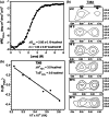
Thermodynamics of the WT‐HlyIIC domain. (a) Urea denaturation of WT‐HlyIIC at 25 C followed by CD mean residue ellipticity (MRE) at 220 nm. (b) Portions of 1H‐15N HSQC spectra of folded WT‐HlyIIC showing that the signal for the trans form of T383 increases at the expense of the cis form with increasing temperature. (c) van 't Hoff plot for the [trans]/[cis] equilibrium in the folded state of WT‐HlyIIC. The van 't Hoff analysis was done using the peak heights for the cis and trans 1H‐15N crosspeaks of residue T383 from (b). Similar analyses for 12 additional residues that give resolved NMR crosspeaks for the cis and trans states in folded WT‐HlyIIC gave consistent results: ∆HFt/Fc = 3.4 ± 0.2 kcal/mol, T∆SFt/Fc = 3.4 ± 0.3 kcal/mol. HlyIIC, C‐terminal domain corresponding to residues D319‐I412 of the hemolysin II from B. cereus; WT, wild type
FIGURE 3
FIGURE 3
Dimerization of P405M‐HlyIIC by NMR (a) 1H‐15N HSQC spectra of P405M showing assigned correlations form the monomer (green) and dimer (purple) states. The spectrum was obtained for a 0.8 mM P405M‐HlyIIC sample in 20 mM NaH2PO4 buffer at pH 6.6, and a temperature of 33 C. The blue arrow indicates the well‐resolved monomer and dimer signals for residue T383. (b) Comparison of 15N transverse relaxation (R2) rates for the HlyIIC‐P405M monomer and dimer. (c) Comparison of translational diffusion constants determined by NMR PFG techniques (see methods) for the HlyIIC‐P405M monomer and dimer. HlyIIC, C‐terminal domain corresponding to residues D319‐I412 of the hemolysin II from B. cereus; PFG, pulsed field gradient
FIGURE 4
FIGURE 4
NMR structure of the P405M‐HlyIIC dimer. (a) NMR structure closest to the ensemble average for the previously reported P405M‐HlyIIC monomer structure. 14 (b) NMR structure closest to average for the P405M‐HlyIIC domain‐swapped dimer structure described in this work (Table 1). Strand β5 (red in the A protomer, purple in the B protomer) is labeled, and the positions of M405, which is substituted for the WT proline in the P405M‐HlyIIC mutant is indicated by white spheres at the Cα carbons. (c) Global superposition of the 25 lowest‐energy NMR conformers to illustrate the precision of the dimer structure. (d) Superposition on the A protomers of the dimer show that while the protomer structures are well defined, the overall dimer structure is less precise due to a limited number of interchain NOEs. The first 11 disordered residues in each structure are not shown. HlyIIC, C‐terminal domain corresponding to residues D319‐I412 of the hemolysin II from B. cereus
FIGURE 5
FIGURE 5
Thermodynamics of HlyIIC dimerization. (a) Plots of dimer versus monomer concentrations for P405M‐HlyIIC determined from the NMR crosspeaks of residue T383 according to Equation (7). The slopes of the data were used to obtain Kd values at each temperature according to Equation (8). (b) van 't Hoff analysis of the temperature‐dependence of Kd using the data from (A). Only the linear part of the data were used, excluding the points at 15 and 35 C. The resulting ∆H and ∆S values are close to those when all temperatures were used (Table 2). (c) Portions of 1H‐15N HSQC spectra of P405M, P405A, and WT‐HlyIIC. Peaks from monomers are labeled green, those from dimer purple, the cis state orange, the trans state cyan, and peaks where conformational states cannot be resolved are labeled black. The downfield peak D* is assigned to the dimer based on its disappearance at low protein concentration. Although a site‐specific assignment for the D* peak is not available, it is well resolved and provides a useful handle of the amount of protein in the dimer state. While the spectra for the P405M and P405A mutants were obtained for 0.8 mM protein concentrations in about 20 min, the spectrum of WT‐HlyIIC was recorded at a 9 mM concentration using a total acquisition time of 13 h. The weak relative intensity of the D* peak attests to the diminished propensity of HlyIIC to dimerize when the WT proline occurs at position 405. HlyIIC, C‐terminal domain corresponding to residues D319‐I412 of the hemolysin II from B. cereus; WT, wild type

Similar articles

Cited by

References

    1. Stenfors Arnesen LP, Fagerlund A, Granum PE. From soil to gut: Bacillus cereus and its food poisoning toxins. FEMS Microbiol Rev. 2008;32:579–606. - PubMed
    1. Turnbull PC, Kramer JM, Jorgensen K, Gilbert RJ, Melling J. Properties and production characteristics of vomiting, diarrheal, and necrotizing toxins of Bacillus cereus . Am J Clin Nutr. 1979;32:219–228. - PubMed
    1. Drobniewski FA. Bacillus cereus and related species. Clin Microbiol Rev. 1993;6:324–338. - PMC - PubMed
    1. Lund T, De Buyser ML, Granum PE. A new cytotoxin from Bacillus cereus that may cause necrotic enteritis. Mol Microbiol. 2000;38:254–261. - PubMed
    1. Andreeva ZI, Nesterenko VF, Yurkov IS, Budarina ZI, Sineva EV, Solonin AS. Purification and cytotoxic properties of Bacillus cereus hemolysin II. Protein Expr Purif. 2006;47:186–193. - PubMed

Publication types

MeSH terms

LinkOut - more resources