. 2021 May;25(10):4596-4607.
doi: 10.1111/jcmm.16253. Epub 2021 Mar 17.

CDX2 enhances natural killer cell-mediated immunotherapy against head and neck squamous cell carcinoma through up-regulating CXCL14

Affiliations

CDX2 enhances natural killer cell-mediated immunotherapy against head and neck squamous cell carcinoma through up-regulating CXCL14

Haitao Wang et al. J Cell Mol Med. 2021 May.

Abstract

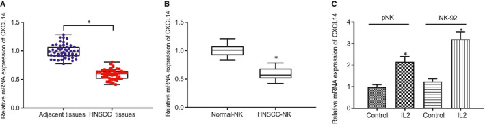
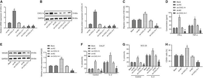
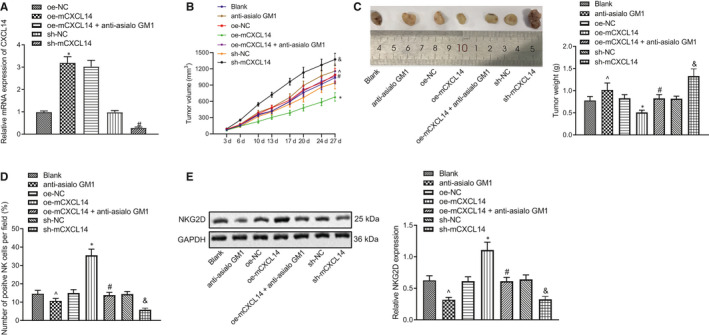
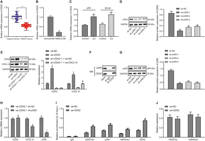
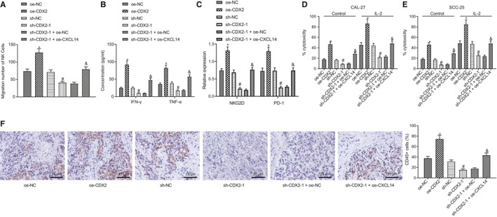
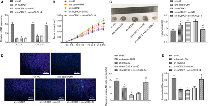
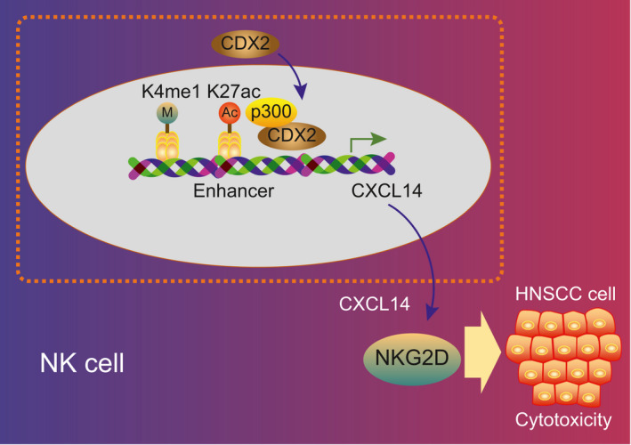
(NK) cells are at the first line of defence against tumours, but their anti-tumour mechanisms are not fully understood. We aimed to investigate the mechanism by which NK cells can mediate immunotherapy against head and neck squamous cell carcinoma (HNSCC). We collected fifty-two pairs of HNSCC tissues and corresponding adjacent normal tissues; analysis by RT-qPCR showed underexpression of CXCL14 in HNSCC tissues. Primary NK cells were then isolated from the peripheral blood of HNSCC patients and healthy donors. CXCL14 was found to be consistently under-expressed in the primary NK cells from the HNSCC patients. However, CXCL14 expression was increased in IL2-activated primary NK cells and NK-92 cells. We next evaluated NK cell migration, IFN-γ and TNF-α expression, cytotoxicity and infiltration in response to CXCL14 overexpression or knockdown using gain- and loss-of-function approach. The results exhibited that CXCL14 overexpression promoted NK cell migration, cytotoxicity and infiltration. Subsequent in vivo experiments revealed that CXCL14 suppressed the growth of HNSCC cells via activation of NK cells. ChIP was applied to study the enrichment of H3K27ac, p300, H3K4me1 and CDX2 in the enhancer region of CXCL14, which showed that CDX2/p300 activated the enhancer of CXCL14 to up-regulate its expression. Rescue experiments demonstrated that CDX2 stimulated NK cell migration, cytotoxicity and infiltration through up-regulating CXCL14. In vivo data further revealed that CDX2 suppressed tumorigenicity of HNSCC cells through enhancement of CXCL14. To conclude, CDX2 promotes CXCL14 expression by activating its enhancer, which promotes NK cell-mediated immunotherapy against HNSCC.

Keywords: CDX2; CXCL14; HNSCC; NK cells; immunotherapy.


Conflict of interest statement

The authors declare that there is no conflict of interest.

Figures

FIGURE 1
FIGURE 1
CXCL14 expression is down‐regulated in head and neck squamous cell carcinoma (HNSCC) and the derived NK cells. (A) RT‐qPCR to detect the expression of CXCL14 in clinical samples of HNSCC patients (n = 52). (B) RT‐qPCR to detect the expression of CXCL14 in NK cells from peripheral blood of healthy volunteers (n = 10) and HNSCC patients (n = 10). (C) RT‐qPCR to detect the expression of CXCL14 in IL‐2‐stimulated NK cells and NK‐92 cells. *P < .05 compared with adjacent normal tissues/normal NK/NK‐92 cells. Measurement data were presented as mean ± standard deviation. Data from tumour and adjacent normal tissues were analysed by paired t test, while data of two groups were compared using unpaired t test. Experiments were repeated 3 times
FIGURE 2
FIGURE 2
CXCL14 enhances NK cell migration and cytolytic activity. (A) RT‐qPCR to detect CXCL14 expression after silencing or overexpressing CLCX14 by lentivirus. (B) Western blot assay to detect the expression of CLCX14 protein after silencing or overexpressing CLCX14 by lentivirus. (C) Transwell to assess the cell migration. (D) ELISA to measure the levels of IFN‐γ and TNF‐α in the medium of NK cells. (E) Western blot assay to analyse the expression of NKG2D in NK cells. (F) The cytotoxicity analysis of primary NK cells to head and neck squamous cell carcinoma (HNSCC) cell line CAL27. (G) The cytotoxicity analysis of primary NK cells to HNSCC cell line SCC‐25. (H) Immunohistochemistry to examine CD45 expression in SCC‐25 tumour spheroid. *P < .05 compared with oe‐NC group, and # P < .05 compared with sh‐NC group. Measurement data were presented as mean ± standard deviation. Data among multiple groups were processed via one‐way ANOVA and Tukey's post hoc test. Experiments were repeated 3 times
FIGURE 3
FIGURE 3
CXCL14 inhibits the growth of head and neck squamous cell carcinoma (HNSCC) in vivo. (A) RT‐qPCR to detect CXCL14 expression in mouse tumours. (B) Measurement of tumour volume. (C) Measurement of tumour weight. (D) IF to detect the NK cell infiltration in mouse tumours. (E) Western blot assay to analyse the expression of NKG2D in mouse tumours. ^P < .05 compared with blank group, *P < .05 compared with oe‐NC group, # P < .05 compared with oe‐mCXCL14 group, and & P < .05 compared with sh‐NC group. Tumours were excised 3 wks after lentivirus injection, and RT‐qPCR, IF and Western blot assay were performed. Measurement data were presented as mean ± standard deviation. Data among multiple groups were compared via one‐way ANOVA and Tukey's post hoc test, and tumour volume data from different time‐points were analysed by repeated measures ANOVA. Experiments were repeated 3 times
FIGURE 4
FIGURE 4
CDX2 increases CXCL14 expression via activating its enhancer in primary NK cells. (A) RT‐qPCR to detect CDX2 expression in clinical samples of head and neck squamous cell carcinoma (HNSCC) patients (n = 52). *P < .05 compared with the adjacent normal tissues. (B) RT‐qPCR to detect CDX2 expression in NK cells of peripheral blood of healthy volunteers and HNSCC patients. *P < .05 compared with NK cells from healthy volunteers. (C) RT‐qPCR to detect CDX2 expression in IL‐2‐stimulated primary NK cells and NK‐92 cells. *P < .05 compared with control NK cells and NK‐92 cells. (D) Western blot assay to analyse the expression of CDX2 after silencing CDX22 in primary NK cells by lentivirus. *P < .05 compared with sh‐NC group. (E) RT‐qPCR and Western blot assay to detect the expression of CDX2, CXCL14, and p300 in NK cells. *P < .05 compared with oe‐NC group, and # P < .05 compared with sh‐CDX2‐1 + oe‐NC group. (F) IP assay to analyse the interaction between p300 and CDX2 in primary NK cells. (G) Western blot assay to analyse the expression of p300 after knocking p300 down in primary NK cells. *P < .05 compared with sh‐NC group. (H) RT‐qPCR to detect the expression of CDX2, CXCL14 and p300. *P < .05 compared with oe‐CDX2 + sh‐NC group. (I) ChIP to analyse the effect of CDX2 on the enrichment of H3K27ac, p300 and H3K4me1 in the enhancer of CLCX14. (J) Western blot assay to analyse the expression of H3K27ac and H3K4me1. *P < .05 compared with oe‐NC group. Measurement data were presented as mean ± standard deviation. Data from tumour and adjacent normal tissues that were normal distributed were analysed by paired t test, while data of two groups were compared using unpaired t test. Data among multiple groups were compared via one‐way ANOVA and Tukey's post hoc test. Experiments were repeated 3 times
FIGURE 5
FIGURE 5
CDX2 enhances NK cell migration and cytolytic activity via up‐regulating CXCL14 expression. (A) Transwell assay to detect the effect of CXCL14 on the migration of NK cells. (B) ELISA to measure the levels of IFN‐γ and TNF‐α in the medium of NK cells. (C) Western blot assay to analyse the expression of NKG2D and PD‐1 in NK cells. (D) The cytotoxicity of NK cells to head and neck squamous cell carcinoma (HNSCC) cell line CAL27. (E) The cytotoxicity of NK cells to HNSCC cell line SCC‐25. (F) Immunohistochemistry to detect the expression of CD45 in SCC‐25 tumour spheroid. *P < .05 compared with oe‐NC, # P < .05 compared with sh‐NC group, and & P < .05 compared with sh‐CDX2 + oe‐NC group. Measurement data were presented as mean ± standard deviation. Data of two groups were compared using unpaired t test. Experiments were repeated 3 times
FIGURE 6
FIGURE 6
CDX2/CXCL14 inhibits the growth of head and neck squamous cell carcinoma (HNSCC) in vivo. (A) RT‐qPCR to detect the expression of CDX2 and CXCL14 in tumours. (B) The measurement of tumour volume. (C) The measurement of tumour weight. (D) IF to detect the NK cell infiltration in tumours. (E) Western blot assay to analyse the expression of NKG2D in mouse tumours. *P < .05 compared with sh‐NC group, and # P < .05 compared with sh‐mCDX2 + oe‐NC group. Tumours were excised 3 wks after lentivirus injection, and RT‐qPCR, IF and Western blot assay were performed. Measurement data were presented as mean ± standard deviation. Data among multiple groups were processed via one‐way ANOVA and Tukey's post hoc test, and tumour volume data from different time‐points were analysed by repeated measures ANOVA. Experiments were repeated 3 times
FIGURE 7
FIGURE 7
CDX2 promotes CXCL14 expression by activating its enhancer, which enhances NK cell–mediated immunotherapy against head and neck squamous cell carcinoma (HNSCC)

Similar articles

Cited by

References

    1. Tsompana M, Gluck C, Sethi I, et al. Reactivation of super‐enhancers by KLF4 in human head and neck squamous cell carcinoma. Oncogene. 2020;39:262‐277. - PubMed
    1. Vigneswaran N, Williams MD. Epidemiologic trends in head and neck cancer and aids in diagnosis. Oral Maxillofac Surg Clin North Am. 2014;26:123‐141. - PMC - PubMed
    1. Tuna M, Amos CI, Mills GB. Genome‐wide analysis of head and neck squamous cell carcinomas reveals HPV, TP53, smoking and alcohol‐related allele‐based acquired uniparental disomy genomic alterations. Neoplasia. 2019;21:197‐205. - PMC - PubMed
    1. Marur S, Forastiere AA. Head and neck squamous cell carcinoma: update on epidemiology, diagnosis, and treatment. Mayo Clin Proc. 2016;91:386‐396. - PubMed
    1. Moreira J, Tobias A, O'Brien MP, et al. Targeted therapy in head and neck cancer: an update on current clinical developments in epidermal growth factor receptor‐targeted therapy and immunotherapies. Drugs. 2017;77:843‐857. - PubMed

MeSH terms